with flowcytometry, cytogenetic and molecular biology findings
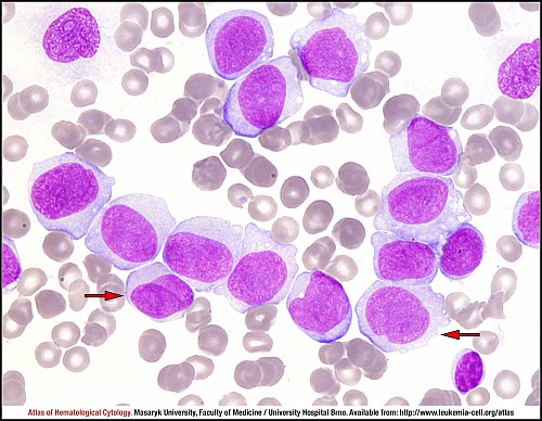
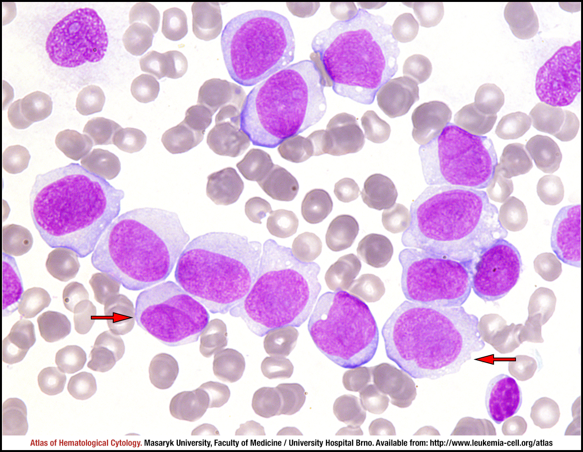
The majority of cells are monoblasts – large cells with abundant cytoplasm, which can be moderately (like in this case) to strongly basophilic; scattered fine azurophilic granules and vacuoles may also be present. Monoblasts usually have round nuclei with delicate lacy chromatin, and one or more large and prominent nucleoli. Promonocytes have more irregular and convoluted nuclear outline (red arrows). In acute monoblastic leukaemia, 80% or more leukaemic cells belong to monocytic lineage, and monoblasts represent the majority of tumour cells (typically ≥ 80%).
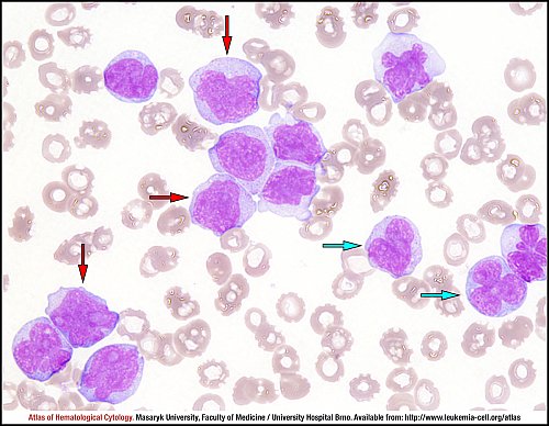
In acute monocytic leukaemia, the majority of tumour cells are promonocytes (red arrows), which are considered as “monoblasts equivalents”, and abnormal monocytes (blue arrows). These monocytes appear immature, yet have a more condensed nuclear chromatin, convoluted or folded nuclei, and more cytoplasmic granulation. Monocytic lineage should be present in 80% or more of all nucleated bone marrow cells; “blast equivalents” (i.e. monoblasts and promonocytes) should reach at least 20%.
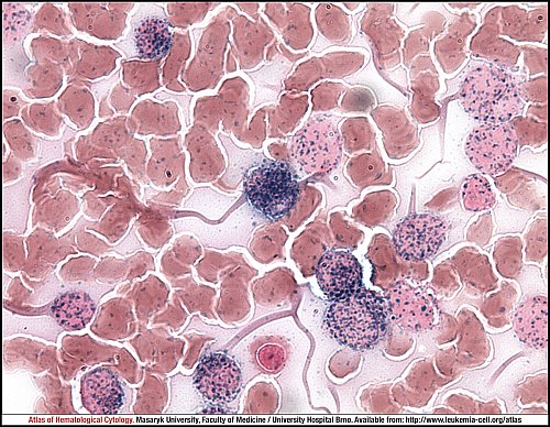
The monocytic nature of leukaemic cells can be visualised by cytochemistry. α-naphthyl acetate esterase staining shows a diffuse cytoplasmic activity in monoblasts, promonocytes and monocytes; this activity is totally or mostly inhibited by natrium fluoride.
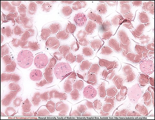
The monocytic nature of leukaemic cells can be visualised by cytochemistry. α-naphthyl acetate esterase staining shows a diffuse cytoplasmic activity in monoblasts, promonocytes and monocytes; this activity is totally or mostly inhibited by natrium fluoride.
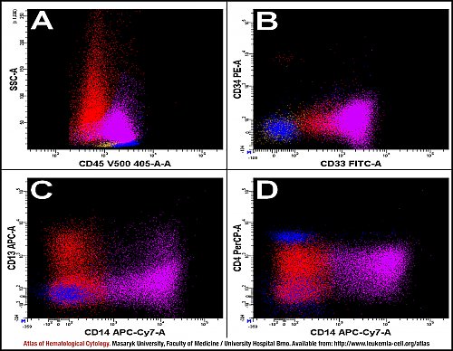
The analysis of bone marrow shows monoblasts (purple), T lymphocytes (blue), B lymphocytes (brown) and granulocytes (red). The population of monoblasts is detected in the region of normal monocytes, but their proportional amount is higher. These cells seem to be almost the same as monocytes, expressing CD45 in variable intensity of SS (A) as well as having a higher expression of CD14 and CD33, a negative expression of CD34 and a low expression of CD4. The negative expression of CD13 is the only characteristics of monoblasts when compared to monocytes (B–D).
Atlas of Haematological Cytology [online]. 2016 [cit. 2024-4-20]. Available from WWW: http://www.leukemia-cell.org/atlas.
2024 CELL - Atlas of Haematological Cytology | site map
 zoom picture
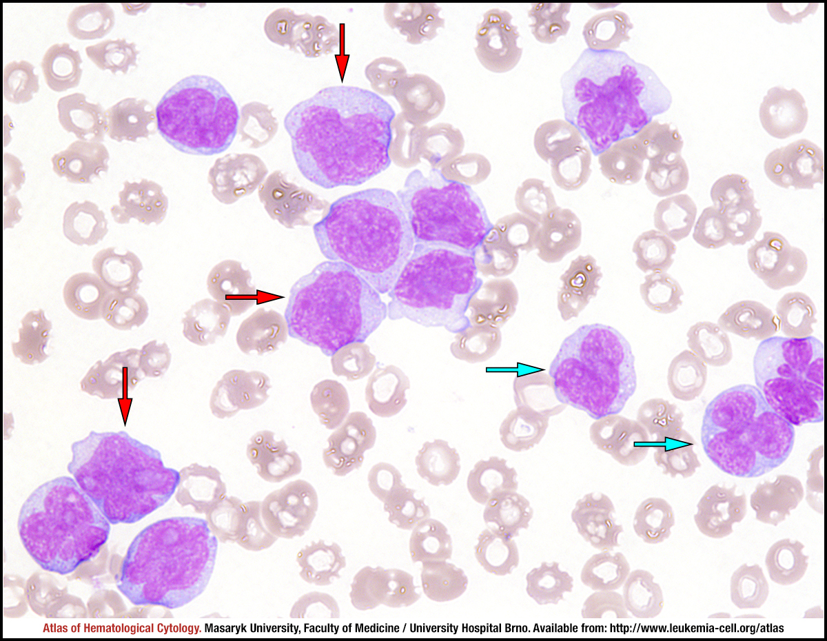 zoom picture
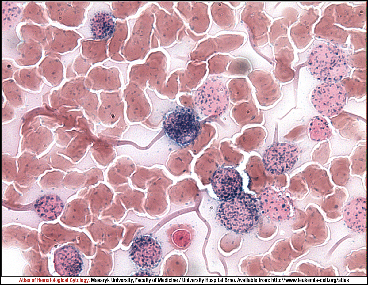 zoom picture
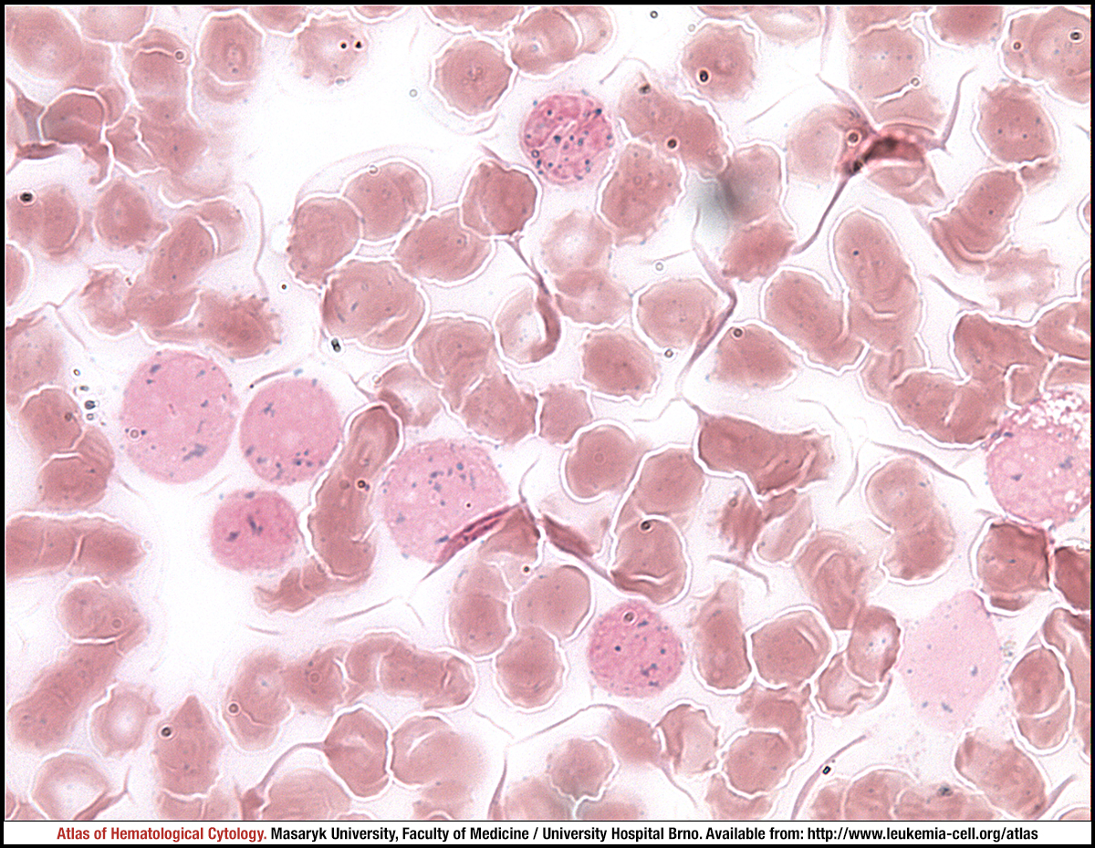 zoom picture
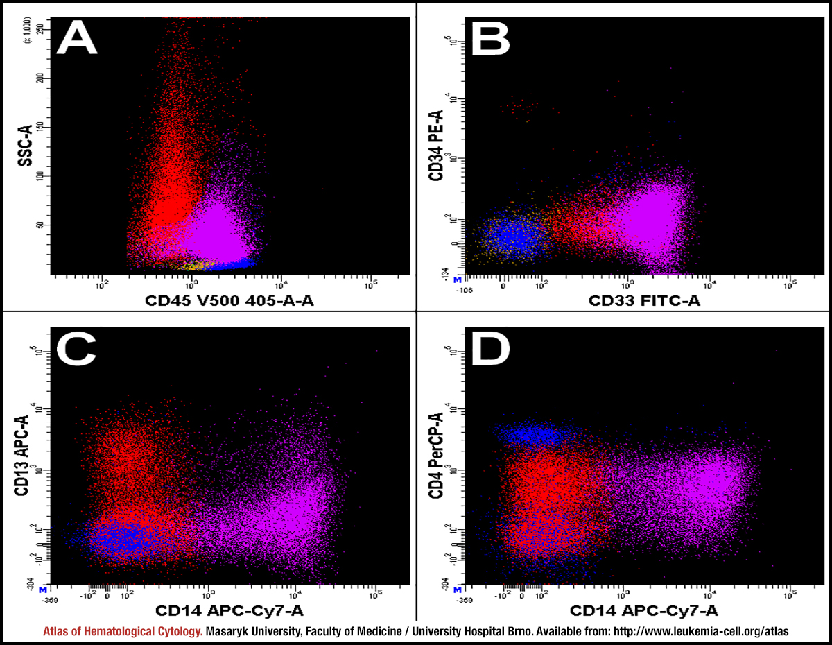 zoom picture