with flowcytometry, cytogenetic and molecular biology findings
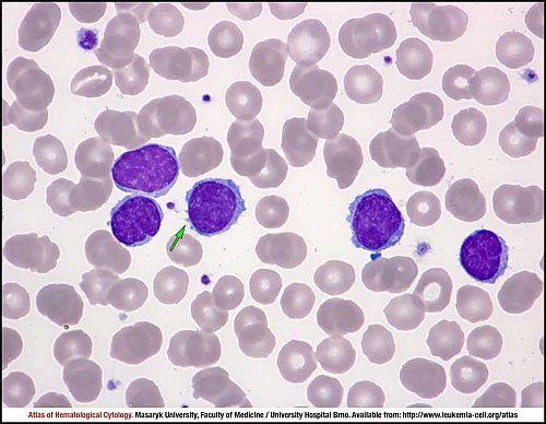
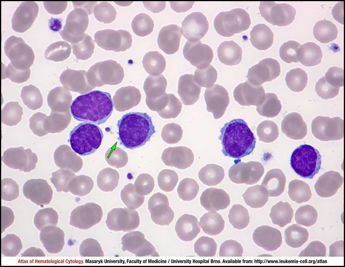
Bone marrow involvement and a leukaemic phase may be present in nodal marginal zone lymphoma. This peripheral blood smear shows centrocyte-like neoplastic lymphocytes. The tumour cells are small to medium-sized with clumped and sometimes irregular nuclear chromatin. The cytoplasm is scanty and basophilic with inconspicuous projections (green arrow). The morphological picture is similar to CLL.
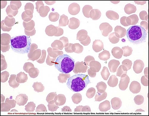
Bone marrow involvement and a leukaemic phase may be present in nodal marginal zone lymphoma. Plasmacytoid morphological features are frequent in the disease. This peripheral blood smear shows neoplastic lymphocytes with plasmacytoid differentiation. The tumour cells have abundant, slightly basophilic cytoplasm and eccentric rounded or oval nuclei with densely clumped chromatin. Sporadically, the nucleolus is prominent (yellow arrow) and perinuclear hof is seen (red arrow). In this case, the distinction from lymphoplasmacytic lymphoma is practically impossible only on the basis of cytology.
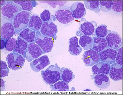
Central nervous system involvement is extremely rare in nodal marginal zone lymphoma. Cytospin of cerebrospinal fluid shows a massive infiltration by monocytoid marginal zone lymphoma cells. The tumour lymphocytes have highly variable irregular shapes of nuclei, moderately clumped chromatin and many prominent nucleoli (yellow arrow). The nuclei are of monocytoid appearance. The cytoplasm is abundant and basophilic, vacuolisation is sporadically observed (red arrow).
Atlas of Haematological Cytology [online]. 2016 [cit. 2024-4-19]. Available from WWW: http://www.leukemia-cell.org/atlas.
2024 CELL - Atlas of Haematological Cytology | site map
 zoom picture
 zoom picture
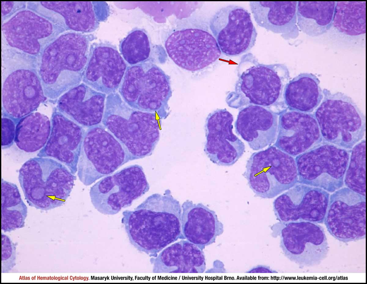 zoom picture