with flowcytometry, cytogenetic and molecular biology findings
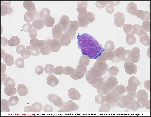
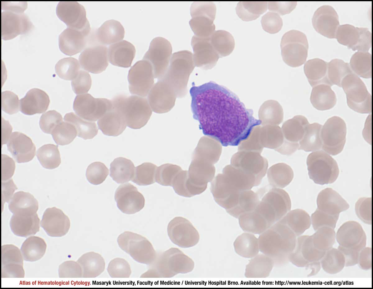
Anaplastic large cell lymphoma cells are uncommon in peripheral blood and this finding is indicative of a worse prognosis. When it occurs, lymphoma cells may be pleomorphic. This particular image shows an undifferentiated blast with a high nuclear-cytoplasmic ratio, fine chromatin, rather deep basophilic cytoplasm and a few clear nucleoli.
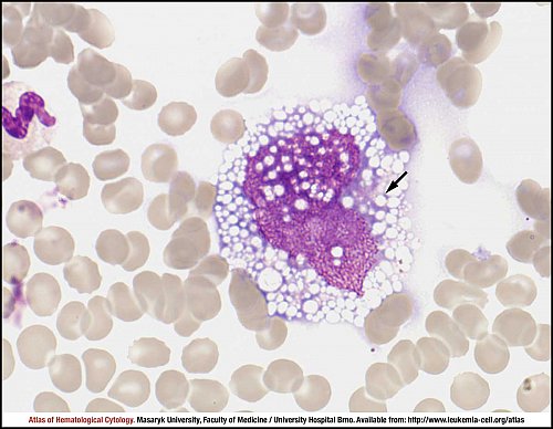
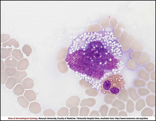
Bone marrow infiltration has been reported in about 1 in 4 (or even more) patients. Lymphoma cells are often infrequent, usually accounting for less than 5% of marrow cells. Neoplastic cells are large and pleomorphic, some being as large as megakaryocytes. They have weakly to strongly basophilic cytoplasm, which may be vacuolated, visibly finely but strongly in this case. A variable proportion of cells meets the criteria of "hallmark cells" in this disease, namely eccentric, horseshoe- or kidney-shaped nuclei often with an eosinophilic region near the nucleus. All of these characteristics are present in this case, only vacuolisation is stronger than usually seen in patients with ALCL, ALK+. The eosinophilic area near the nucleus is present as usual, but hardly visible due to a strong vacuolisation; however, it is still detectable in this case (black arrow).
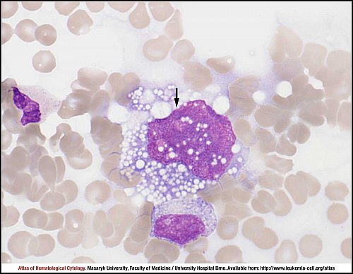
Another example of ALCL ALK+ tumour cell. The cell has kidney-shaped nucleus, fine nuclear chromatin pattern with hardly visible ring-like nucleoli (black arrow) and a strongly vacuolated cytoplasm.
Occasional phagocytic tumour cells can occur in this lymphoma. The phagocytosis of erythrocytes is present in this case - three of them are clear, the fourth one is almost destroyed (in the middle). Associated macrophage proliferation and haemophagocytosis are common and they can may lead to peripheral blood cell cytopenia (not shown in this image).
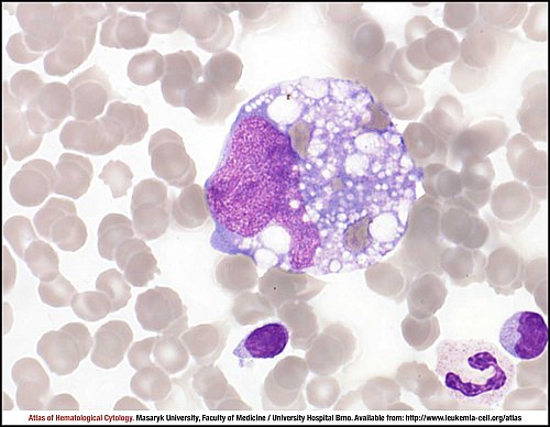
The tumour cell has an irregular nuclear outline, a coarse open chromatin pattern and multiple prominent dark nucleoli (black arrows).
This imprint of lymph node shows a massive involvement by anaplastic large cell lymphoma. The size and appearance of tumour cells is variable. The neoplastic lymphocytes are very large to monstrous (black arrow) and pleomorphic. The cytoplasm is abundant, strongly basophilic and often vacuolised (green arrows). Nuclear chromatin is very fine and irregular, multiple bluish nucleoli are prominent (red arrows). A sporadic eosinophilic leucocyte is admixed (yellow arrow).
Atlas of Haematological Cytology [online]. 2016 [cit. 2024-4-25]. Available from WWW: http://www.leukemia-cell.org/atlas.
2024 CELL - Atlas of Haematological Cytology | site map
 zoom picture
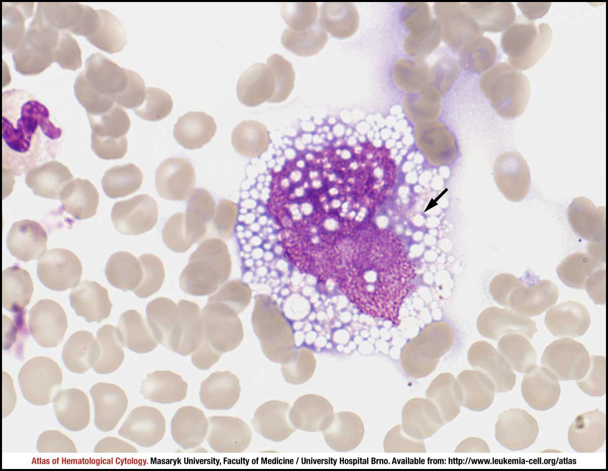 zoom picture
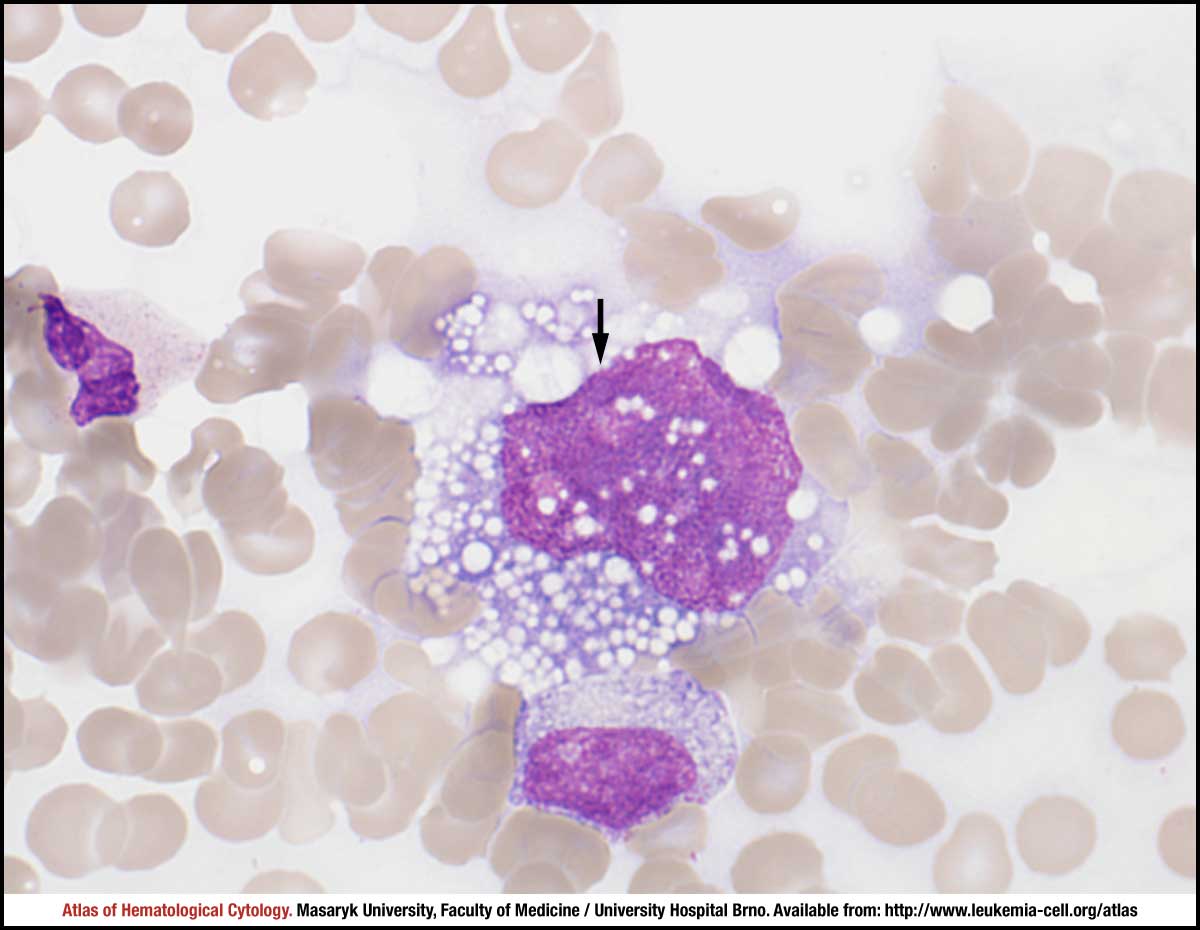 zoom picture
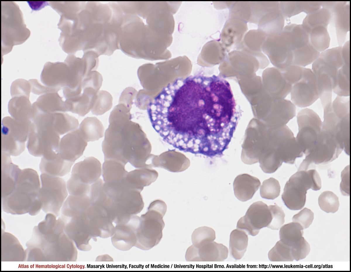 zoom picture
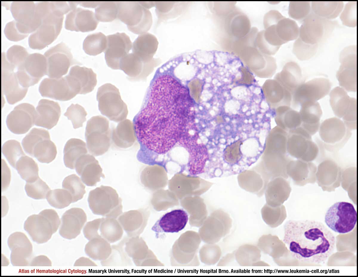 zoom picture
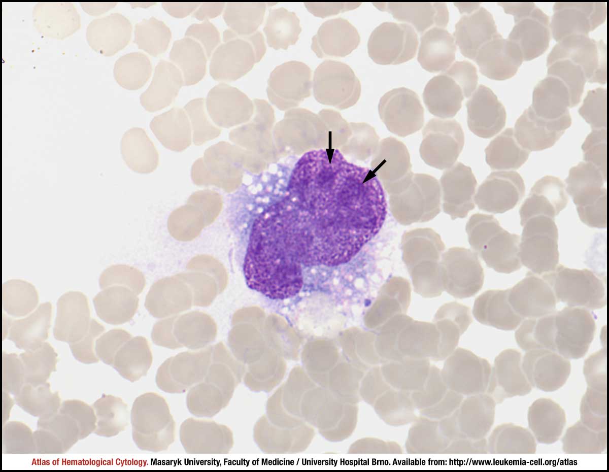 zoom picture
 zoom picture
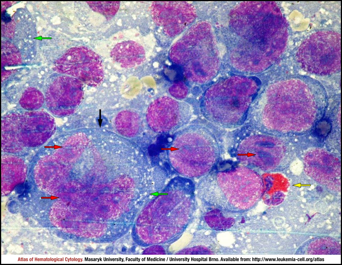 zoom picture