with flowcytometry, cytogenetic and molecular biology findings
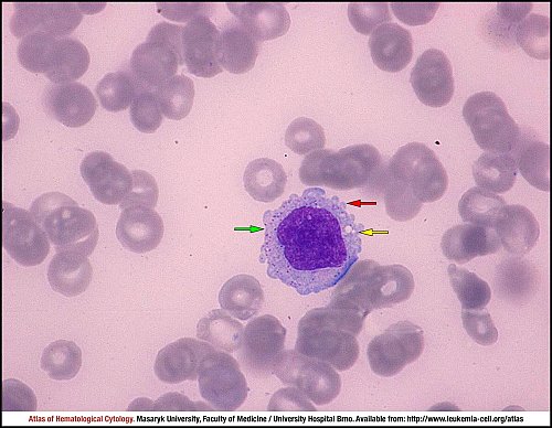
This bone marrow smear shows a neoplastic lymphocyte in hepatosplenic T-cell lymphoma. The neoplastic cell is large, the abundant slightly basophilic cytoplasm has marginal irregularities (red arrow), some vacuoles (yellow arrow) and fine azurophilic cytotoxic granules (green arrow). The nuclear chromatin is loosely condensed.
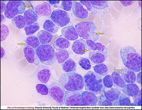
Nodal involvement is extremely rare in hepatosplenic T-cell lymphoma. This lymph node imprint shows an infiltration of pleomorphic small to large lymphocytes. The neoplastic cells have condensed nuclear chromatin without nucleoli and abundant basophilic cytoplasm with fine azurophilic cytotoxic granules (yellow arrows).
Atlas of Haematological Cytology [online]. 2016 [cit. 2024-4-26]. Available from WWW: http://www.leukemia-cell.org/atlas.
2024 CELL - Atlas of Haematological Cytology | site map
 zoom picture
 zoom picture