with flowcytometry, cytogenetic and molecular biology findings
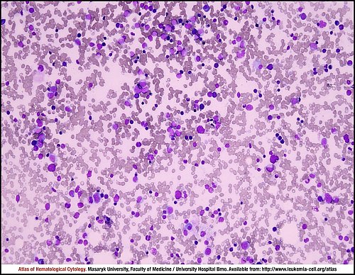
Marrow aspirate smear with an adequate cellularity and an almost proportional representation of haematopoietic cells.
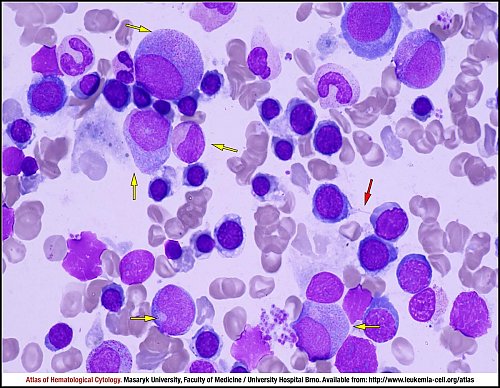
Numerous dysplastic erythroblasts with dehaemoglobinised cytoplasm, some of them containing pyknotic nuclei. On the right, two cells are connected by a cytoplasmic bridge (red arrow). Many granulocytes (yellow arrows) are anisocytic, showing hypogranularity or dysgranularity of cytoplasm and signs of nuclear-cytoplasmic asynchrony.
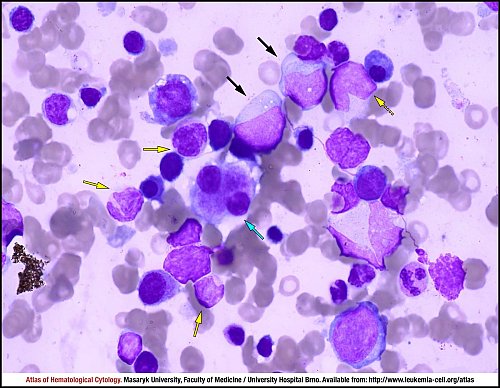
Four dysplastic neutrophilic mature granulocytes, which are microcytic and contain bizarrely segmented nuclei, are marked by yellow arrows. The dotted arrow indicates a megaloblastoid metamyelocyte with agranular cytoplasm, whereas black arrows indicate two larger myeloid blasts without granules. A small dysplastic megakaryocyte with two nuclei is seen in the centre (cyan arrow).
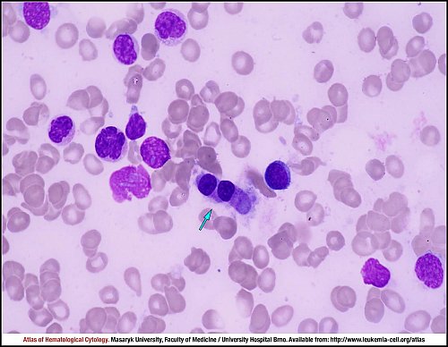
According to diagnostic criteria for MDS-RS, myeloblasts (black arrows) must not exceed 5% of the nucleated bone marrow cells and blasts must not contain Auer rods in their cytoplasm. Furthermore, the image shows two hyposegmented mature neutrophils (dotted yellow arrows) and one pseudopelgeroid neutrophil (yellow arrow). The lower right part of the image shows six predominantly basophilic erythroblasts with impaired haemoglobinisation of cytoplasm, which is an indication of dyserythropoiesis.
A dysplastic megakaryocyte with two nuclei is indicated by a cyan arrow in the centre of the image.
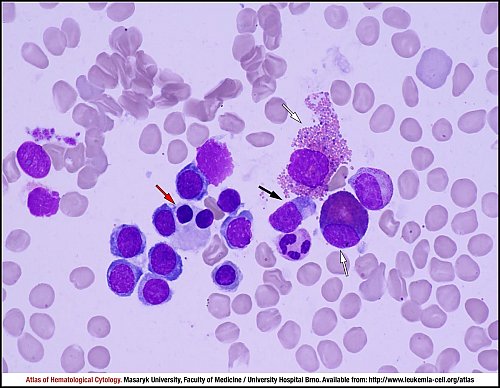
The binuclear macrocytic polychromatophilic erythroblast with partially pyknotic nuclei (red arrow) is dysplastic. A myeloblast is indicated by a black arrow. White arrows indicate eosinophilic myelocytes: the upper cell is already disintegrating.
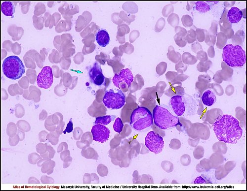
A typical small micromegakaryocyte is indicated by a cyan arrow. Dysplastic neutrophils are indicated by yellow arrows: a binuclear metamyelocyte (dotted arrow), a small myelocyte containing almost no cytoplasm further on the right (dashed arrow) and a bizarrely segmented mature neutrophil on the very right (full arrow). The black arrow indicates a myeloblast.
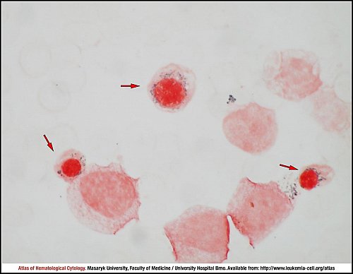
Iron-stained marrow aspirate smear with three ring sideroblasts (red arrows), which are hallmarks of dyserythropoiesis.
Atlas of Haematological Cytology [online]. 2016 [cit. 2026-5-12]. Available from WWW: http://www.leukemia-cell.org/atlas.
2026 CELL - Atlas of Haematological Cytology | site map
 zoom picture
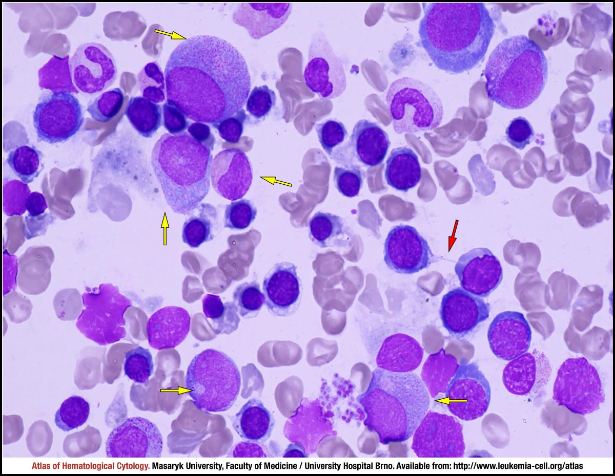 zoom picture
 zoom picture
 zoom picture
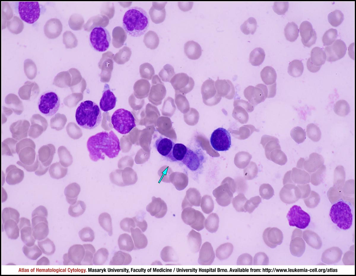 zoom picture
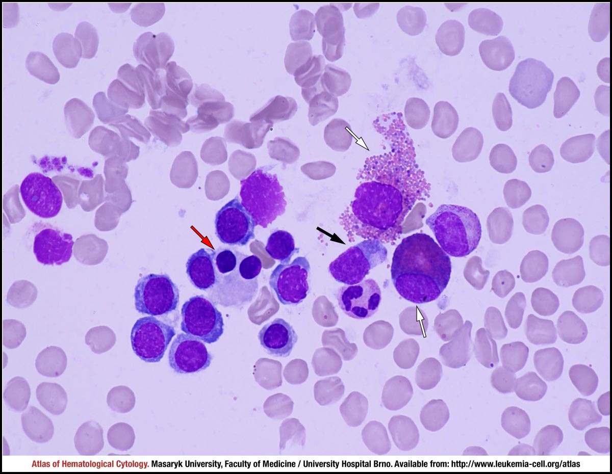 zoom picture
 zoom picture
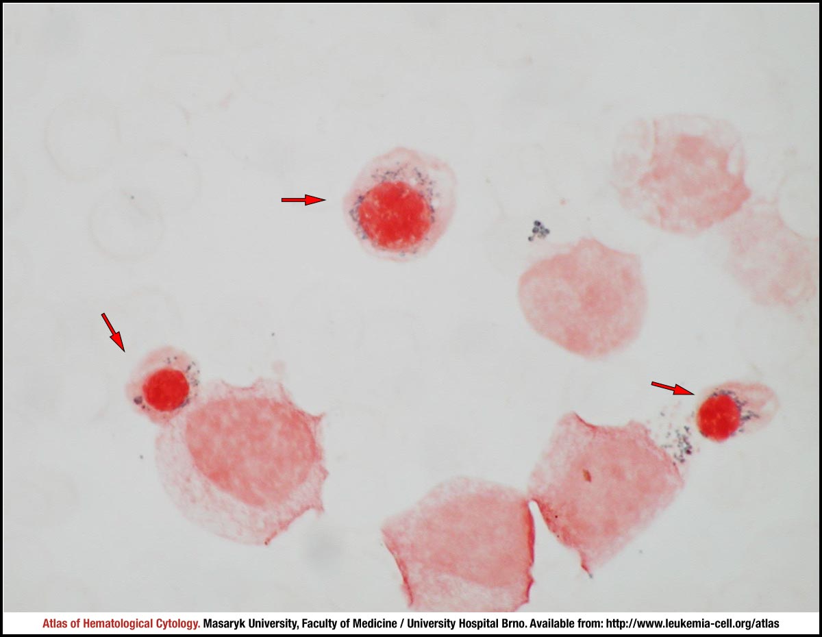 zoom picture