with flowcytometry, cytogenetic and molecular biology findings
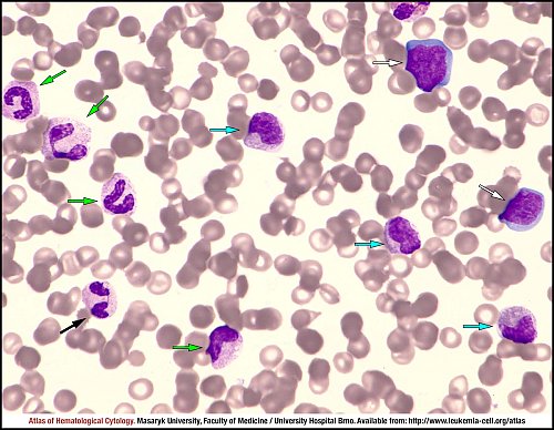
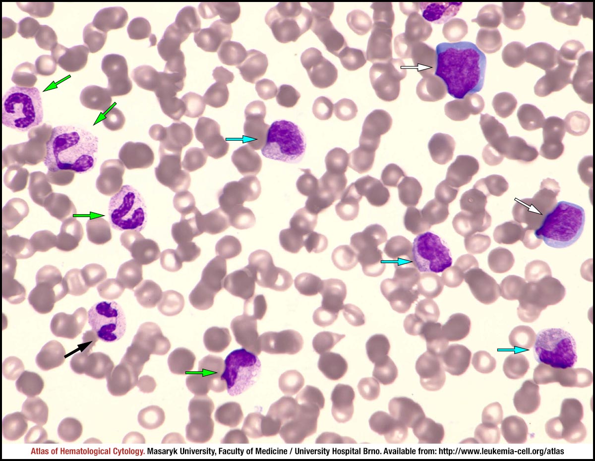
Peripheral blood smear showing neutrophilia with a left shift – neutrophilic cells at different stages of maturation – metamyelocytes (blue arrows), bands (green arrows) and one segmented neutrophil (black arrow). Some of granulocytes are hypogranular and with vacualisation; there are two lymphoblasts (white arrows) with a diameter of about 16–18 µm, showing a nucleus with dispersed chromatin pattern, some nucleoli and deep blue cytoplasm.
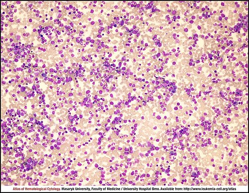
Mild hypercellular marrow aspirate smear with predominant granulopoiesis.
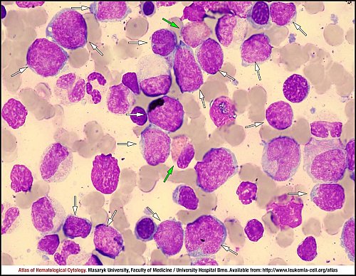
Marrow aspirate smear: many pleomorphic lymphoblasts (white arrows) varying from small blasts with scant cytoplasm, condensed chromatin and indistict nucleoli to larger cells with moderate amounts of blue cytoplasm, occasionally vacuolated. Other cells are from granulocytic lineage – neutrophils and eosinophils (green arrows).
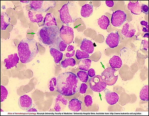
Marrow aspirate smear: many pleomorphic lymphoblasts, cells from granulocytic lineage and four eosinophils (green arrows) of varying maturity.
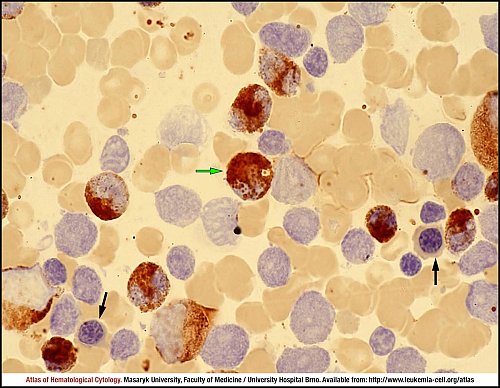
Marrow aspirate smear: erythroblasts (black arrows) and many lymphoblasts are negative, whereas neutrophils and one eosinophil (green arrow) are positive.
Atlas of Haematological Cytology [online]. 2016 [cit. 2025-7-08]. Available from WWW: http://www.leukemia-cell.org/atlas.
2025 CELL - Atlas of Haematological Cytology | site map
 zoom picture
 zoom picture
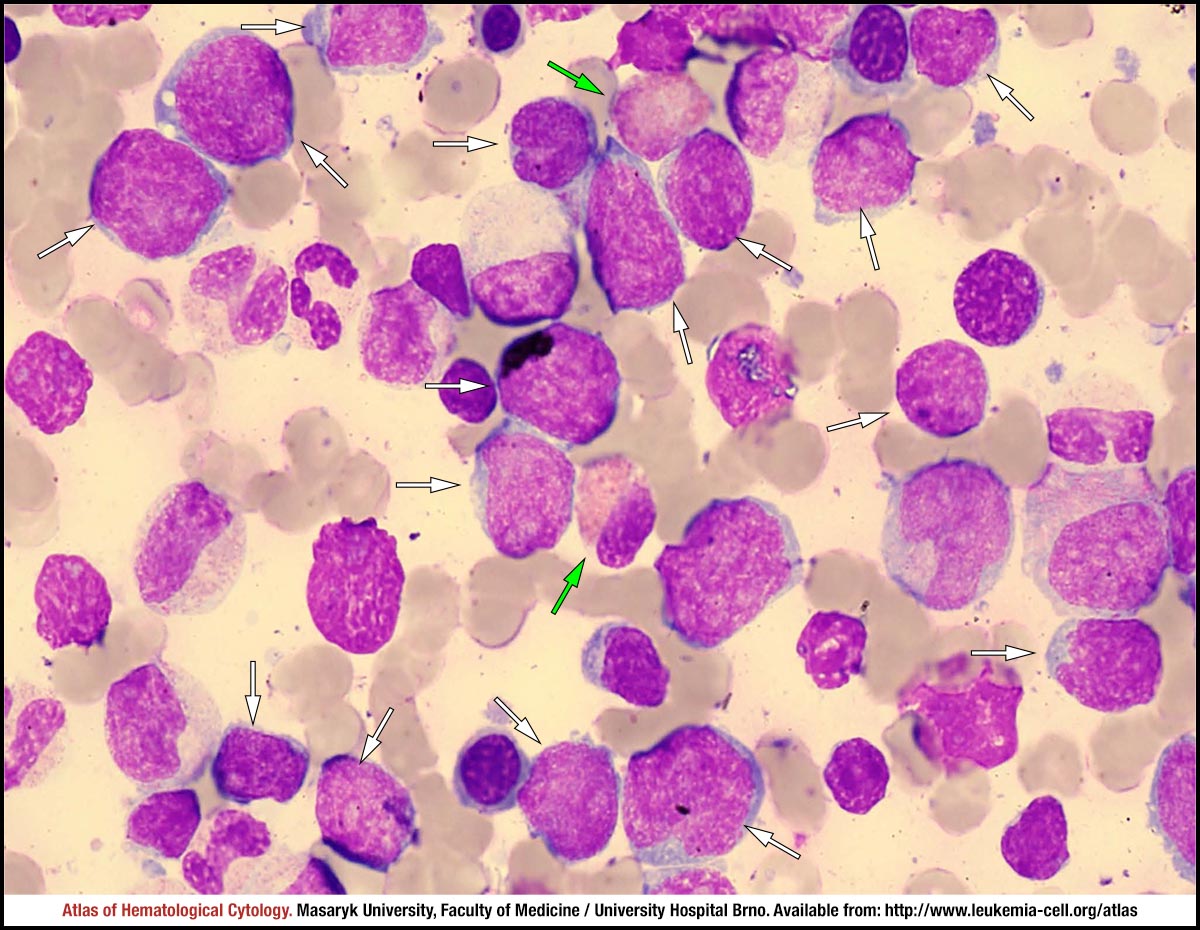 zoom picture
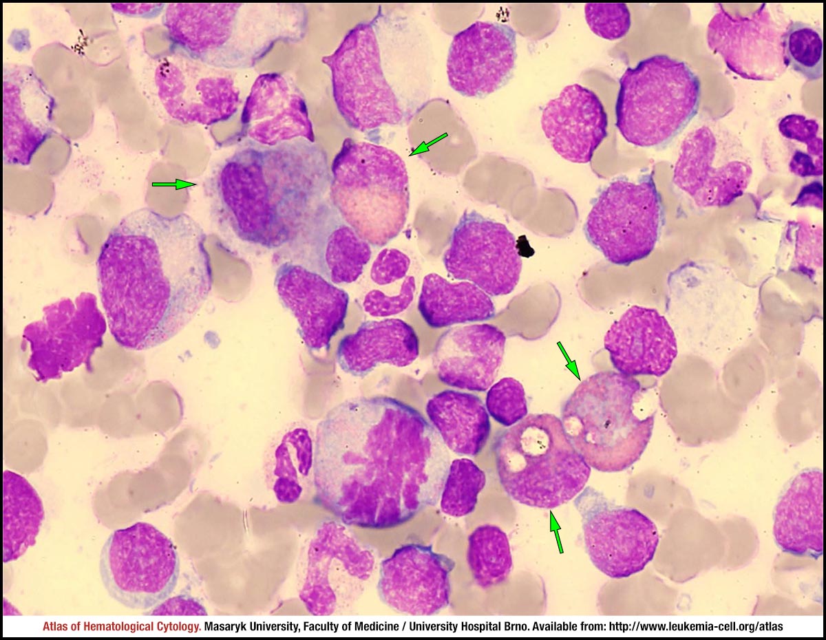 zoom picture
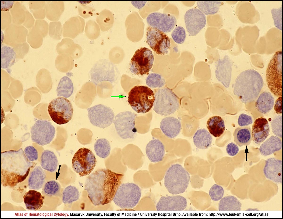 zoom picture