with flowcytometry, cytogenetic and molecular biology findings
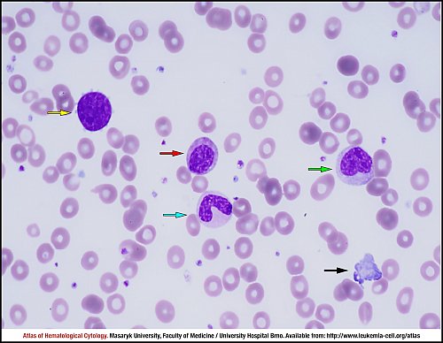
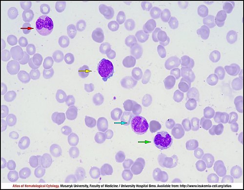
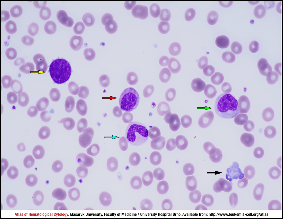
Peripheral blood smear with a left shift: bare nuclei of a megakaryocyte with the rest of cytoplasm (yellow arrow), a myelocyte (red arrow), a band (blue arrow), a metamyelocyte (green arrow) and a fragment of megakaryocytic cytoplasm (black arrow).
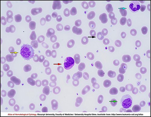
Peripheral blood smear with a left shift: a promyelocyte (yellow arrow), a basophil (red arrow), a segment (blue arrow), a hypogranular myelocyte (green arrow) and basophilic stippling in an erythrocyte (black arrow).
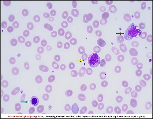
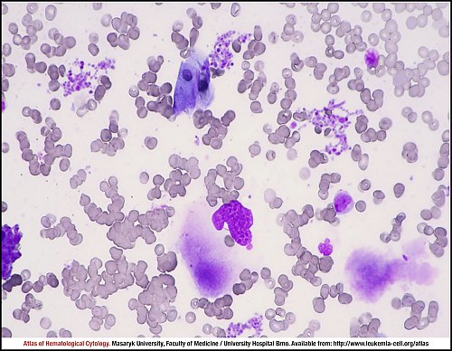
Peripheral blood smear with a left shift: a myelocyte (yellow arrow), a monocyte (red arrow) and an erythroblast (blue arrow).
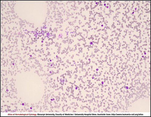
Hypocellular marrow aspirate smear with clumps of thrombocytes.
Hypocellular marrow aspirate smear with clumps of thrombocytes, fragments of a megakaryocyte and cytoplasm.
Hypocellular marrow aspirate smear with peripheral blood cells: a blast (yellow arrow), a band (red arrow), a metamyelocyte (blue arrow) and a monocyte (green arrow).
Primary myelofibrosis, overt fibrosis. 64-year old woman, left-shifted leucocytosis, thrombocytosis over 1,000×109/L. Extremely increased and clustering megakaryocytes with conspicuous “dysplastic” features and emperipolesis.
Bone marrow trephine biopsy, haematoxylin and eosin stain.
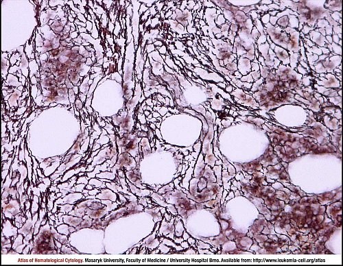
Primary myelofibrosis, overt fibrosis. 72-year old man, leucocytosis 16×109/L, thrombocytosis 767×109/L, JAK2 unmutated. Intense reticulin fibrosis (MF-2 according to the EUMNET classification).
Bone marrow trephine biopsy, Gomori silver impregnation stain.
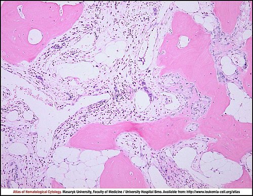
Primary myelofibrosis, overt fibrosis/osteomyelosclerosis. 67-year old woman, progressing leucocytosis up to 30.5×10⁹/L, BCR/ABL negative. Increased, irregular and thickened bone trabecules with new bone formation and osteoblastic rims. Intertrabecular spaces are partially fibrotised with a presence of collagen fibres (MF-3 according to the EUMNET classification).
The overall cellularity is already low and there is a dense cluster of “dysplastic” megakaryocytes.
Bone marrow trephine biopsy, haematoxylin and eosin stain.
Atlas of Haematological Cytology [online]. 2016 [cit. 2026-5-14]. Available from WWW: http://www.leukemia-cell.org/atlas.
2026 CELL - Atlas of Haematological Cytology | site map
 zoom picture
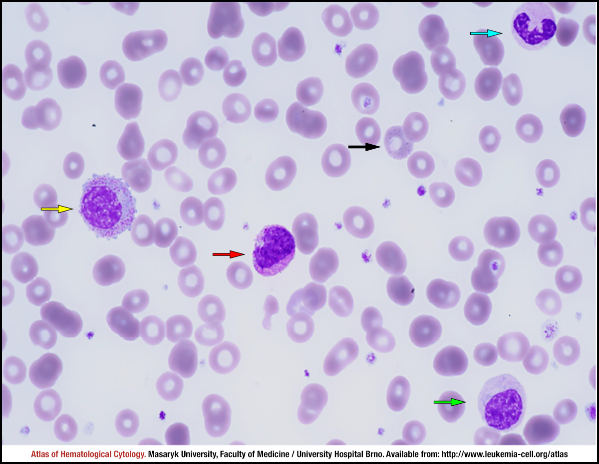 zoom picture
 zoom picture
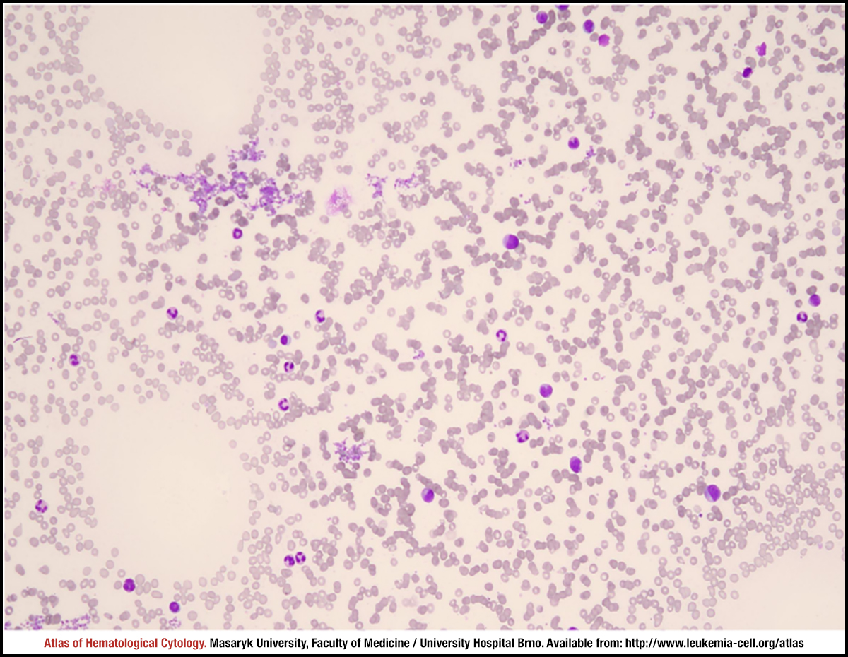 zoom picture
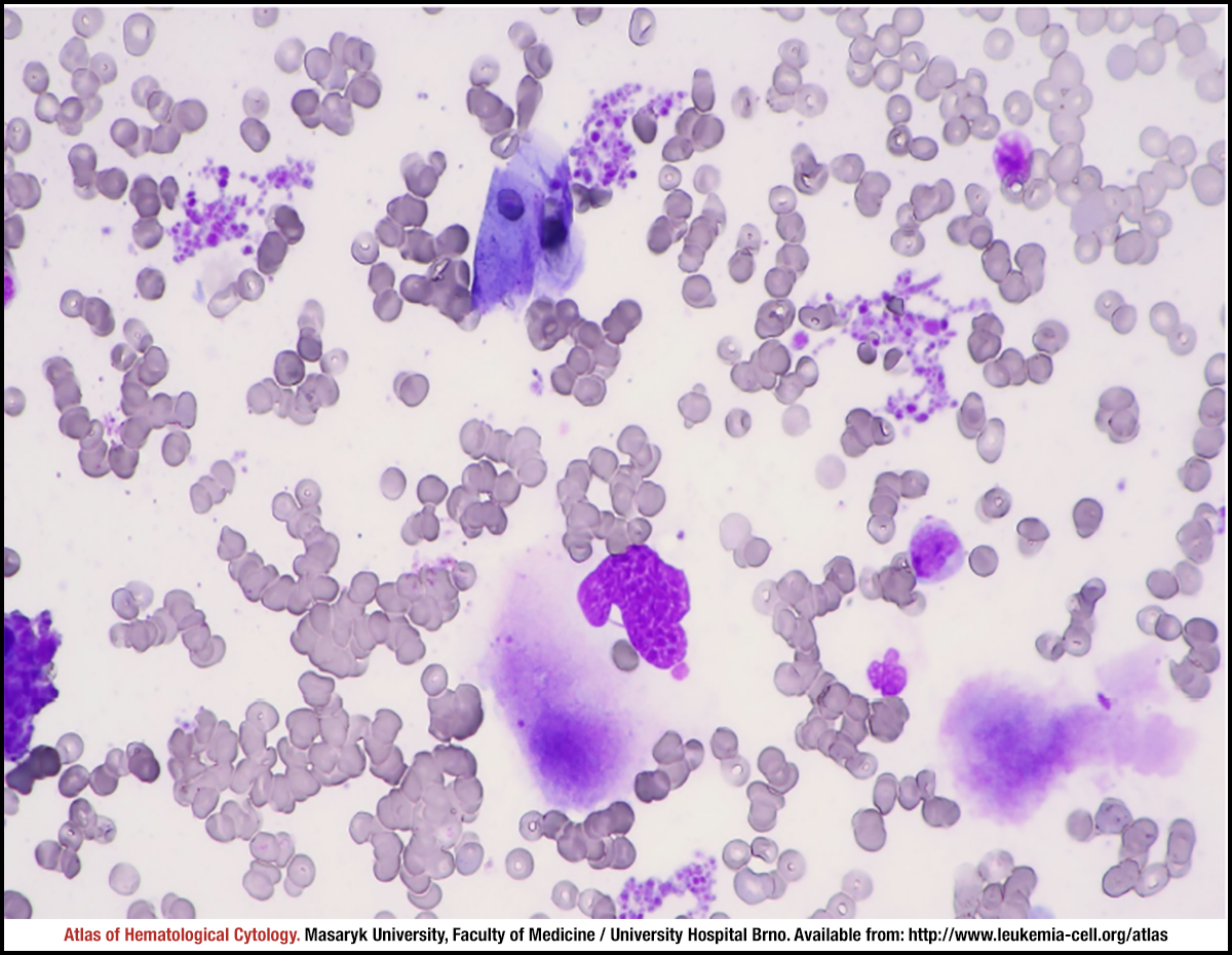 zoom picture
 zoom picture
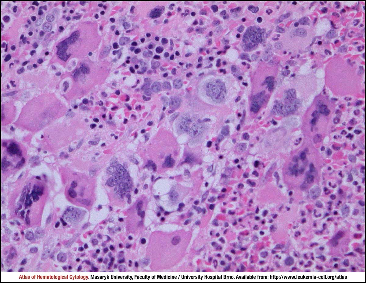 zoom picture
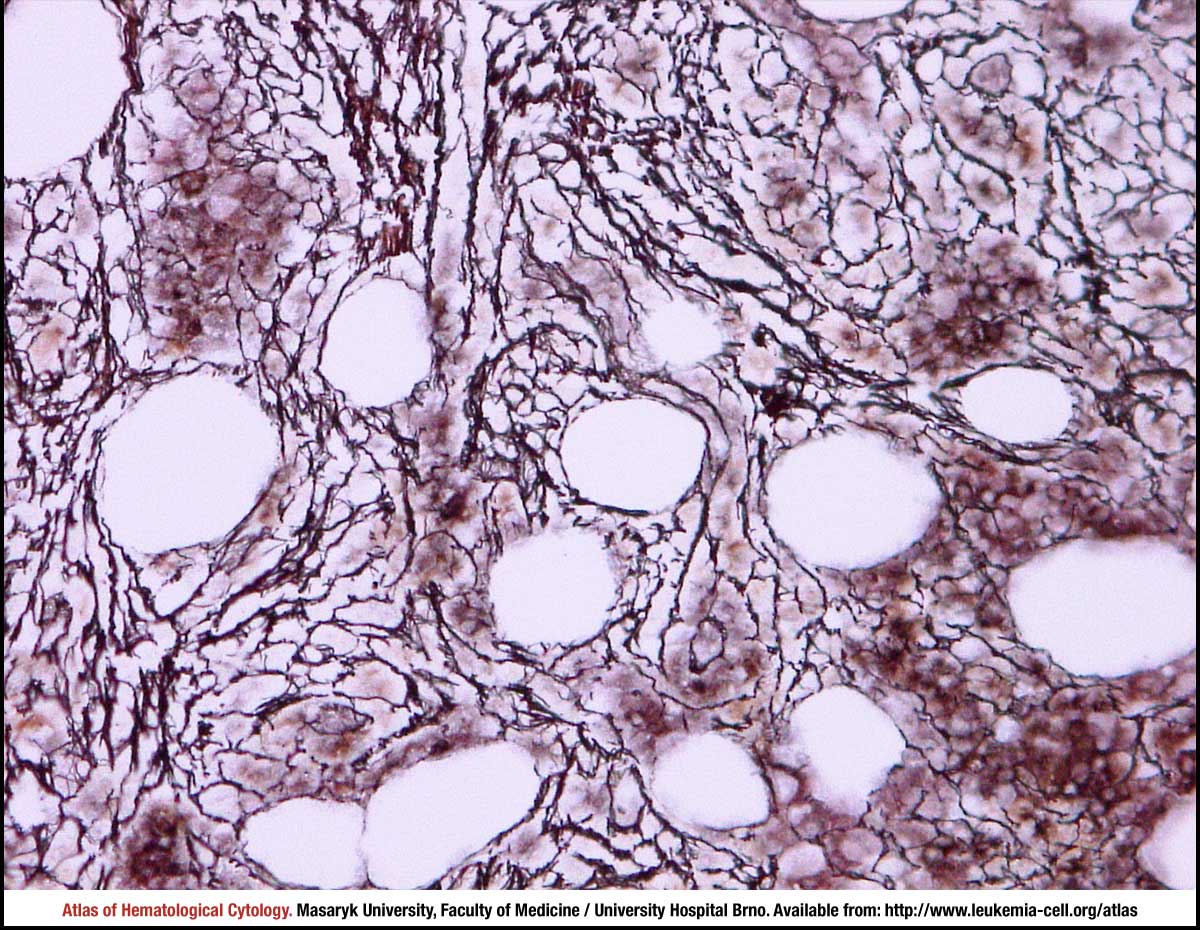 zoom picture
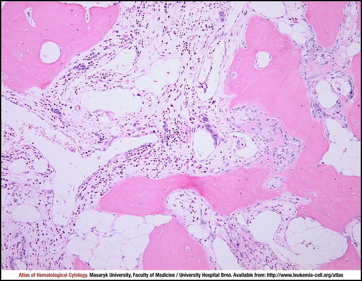 zoom picture