with flowcytometry, cytogenetic and molecular biology findings
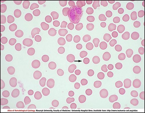
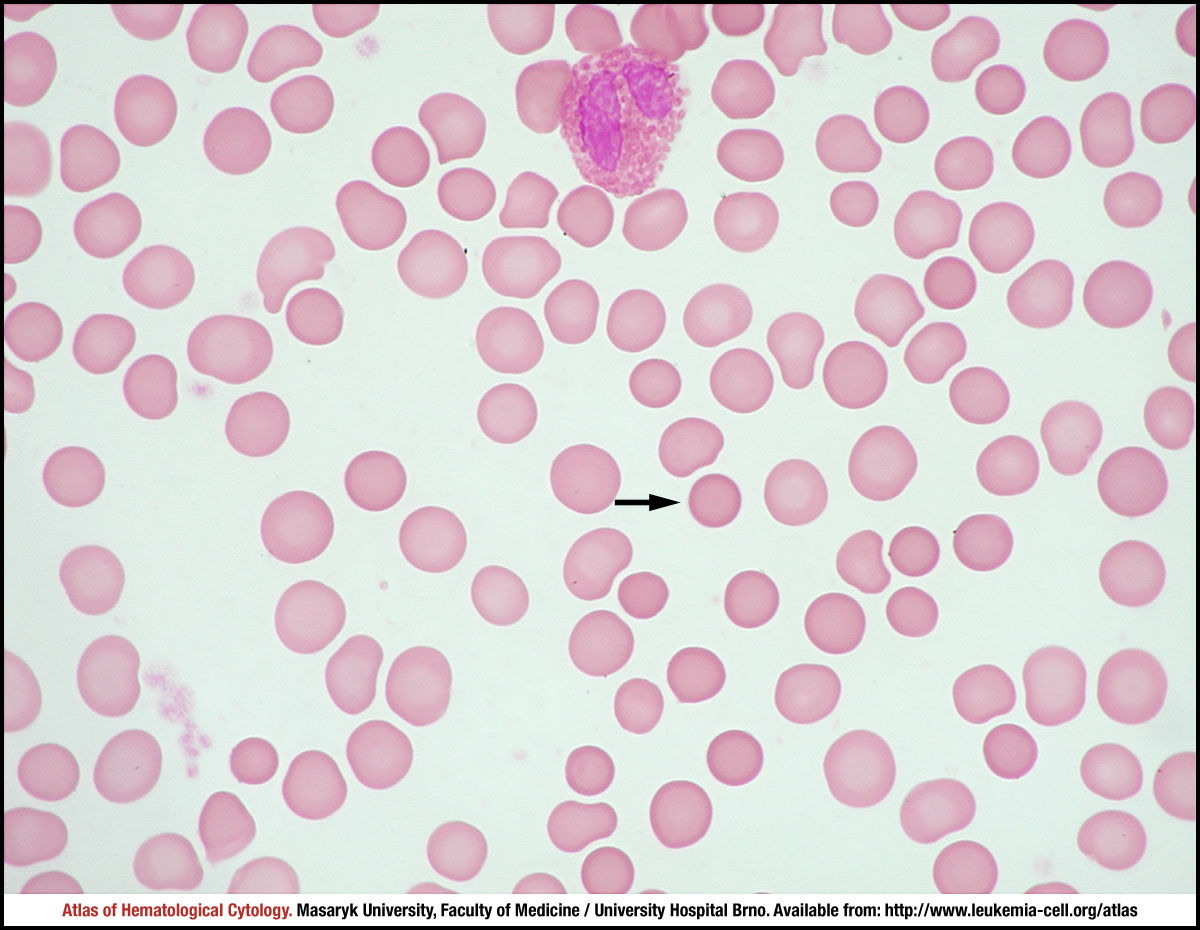
A segmented eosinophil is shown on the top of the image. Numerous spherocytes, i.e. small hyperchromic erythrocytes, typically with no central pallor, are shown mainly in the right part of the image; one of them is marked by a black arrow.
Atlas of Haematological Cytology [online]. 2016 [cit. 2024-4-20]. Available from WWW: http://www.leukemia-cell.org/atlas.
2024 CELL - Atlas of Haematological Cytology | site map
 zoom picture