with flowcytometry, cytogenetic and molecular biology findings
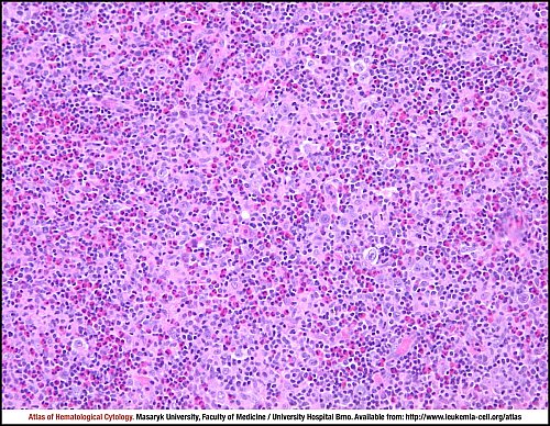
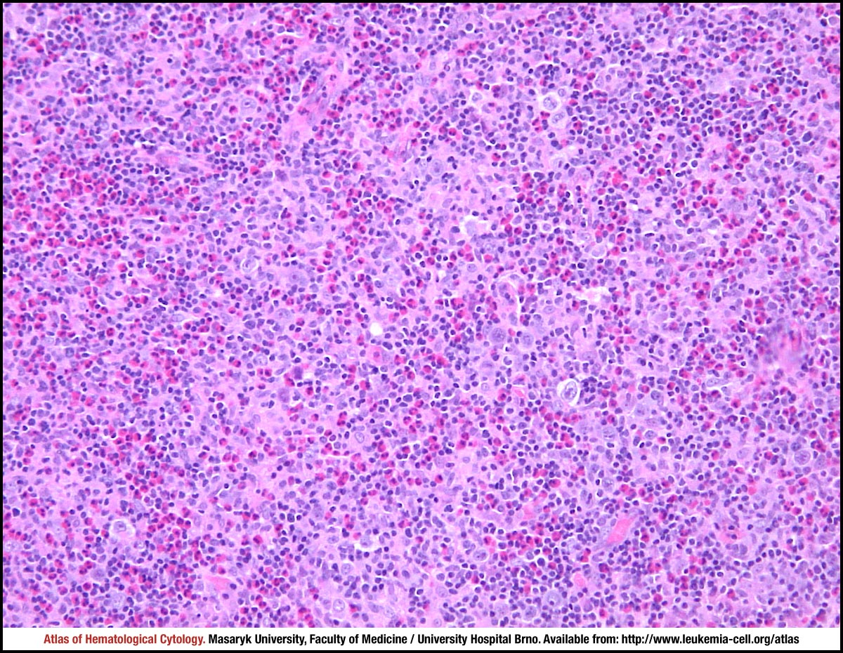
The structure of the lymph node is almost totally effaced by the paracortex expanded by a tumour. Several large atypical cells and a very extensive admixture of eosinophilic leucocytes can be seen even in this low-power view.
Lymph node biopsy, haematoxylin and eosin stain.
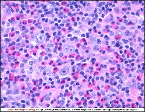
The disperse large atypical Hodgkin/Reed-Sternberg (HRS) cells are present, one of them is binuclear with mirror-imaged vesicular nuclei and inclusion-like nucleoli, the features typical for diagnostic RS cell. The rich “inflammatory” background consists of small lymphocytes, plasma cells, disperse macrophages and a lot of eosinophils.
Lymph node biopsy, haematoxylin and eosin stain.
Atlas of Haematological Cytology [online]. 2016 [cit. 2026-7-11]. Available from WWW: http://www.leukemia-cell.org/atlas.
2026 CELL - Atlas of Haematological Cytology | site map
 zoom picture
 zoom picture