with flowcytometry, cytogenetic and molecular biology findings
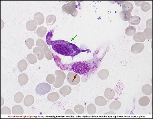
This picture of bone marrow smear shows two tumour cells in a case of systemic mastocytosis. Both cells are atypical mastocytes – “atypical I” according to classification by Sperr at al.1. The first atypical mastocyte (green arrow) has an oval nucleus and cytoplasmatic elongations when compared to normal mastocytes, which usually have a centrally located round nucleus and a regular outline. The second atypical mastocyte (red arrow) is a typical spindle-shaped hypogranular mastocyte, which is one of the most important cytomorphological characteristics of systemic mastocytosis (also called atypical I mastocyte). The aspirate smear must be examined for the presence, numbers (percentage) and morphology of mast cells, and this analysis is to be carried out at a fair distance from any bone marrow particles or larger cell aggregates for the diagnosis of systemic mastocytosis.
1 Sperr WR, Escribano L, et al. Morphologic properties of neoplastic mast cells: delineation of stages of maturation and implication for cytological grading of mastocytosis. Leuk Res 2001; 25(7): 529-536.
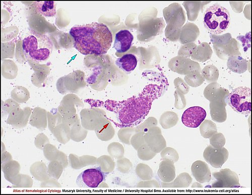
The hypogranulated spindle-shaped mastocyte (“atypical I mastocyte” according to Sperr WR et al.1; red arrow) is the most typical cell found in cases of systemic mastocytosis. Cells like this one, together with normally round mastocytes with centrally located round nuclei and with a typical “mast cells’” granularity are the most frequently found cells in cases of the so-called well-defined systemic mastocytosis (WDSM). An increased granulopoiesis may involve neutrophil, eosinophil (blue arrow) and basophil lineages.
1 Sperr WR, Escribano L, et al. Morphologic properties of neoplastic mast cells: delineation of stages of maturation and implication for cytological grading of mastocytosis. Leuk Res 2001; 25(7): 529-536.
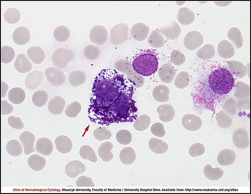
The so-called “atypical II mastocyte” according to Sperr WR et al.1 (red arrow) is another morphologically recognisable tumour cell in cases of systemic mastocytosis. This cell has a bilobed nucleus which is analogous to promastocytes when mast cells are cultured in vitro. Damaged mast cells can also be seen.
1 Sperr WR, Escribano L, et al. Morphologic properties of neoplastic mast cells: delineation of stages of maturation and implication for cytological grading of mastocytosis. Leuk Res 2001; 25(7): 529-536.
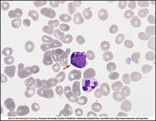
The so-called “atypical III mastocyte” according to Sperr WR et al.1 is the third type of cell which can occur in cases of systemic mastocytosis; it is a blast cell with metachromatically stained granules (red arrow). Bone marrow cells count and the number of different types of mast cells (typical and atypical I – III) have a prognostic significance for the disease course. The presence of more than 5% mastocytes in bone marrow films or having more than 10% or more atypical cells (atypical II mastocytes presented in the previous picture or atypical III mastocytes) among mast cells correlates with a worse prognosis. Nevertheless, the presence of less than 5% of mast cell in bone marrow smears does not preclude heavy mast cell infiltration, which must be demonstrated histologically.
1 Sperr WR, Escribano L, et al. Morphologic properties of neoplastic mast cells: delineation of stages of maturation and implication for cytological grading of mastocytosis. Leuk Res 2001; 25(7): 529-536.
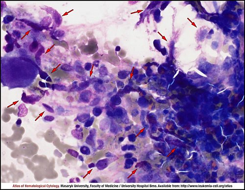
The bone marrow aspirate can contain bone marrow fragments or cell clusters, which also involve numerous neoplastic mast cells. Most of them are atypical spindle-shaped tumour cells (red arrows); however, some of them look like normal mastocytes (green arrow), i.e. oval cells with centrally located non-lobular nuclei and cytoplasm packed with purple granules.
The presence of infiltrates of mast cells, which contain more than 15 tumour cells, is the major criterion for the histologic diagnosis. These large aggregates can be rarely found in bone marrow aspirate smears. Tumour cells are easily identifiable due to their spindle-like shapes and purple granulation (red arrows) in these bone marrow fragments.
Atlas of Haematological Cytology [online]. 2016 [cit. 2026-6-25]. Available from WWW: http://www.leukemia-cell.org/atlas.
2026 CELL - Atlas of Haematological Cytology | site map
 zoom picture
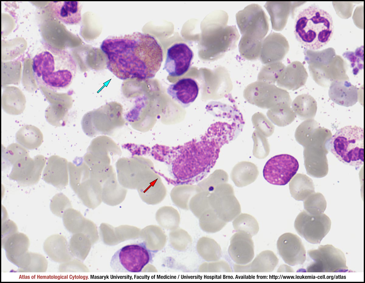 zoom picture
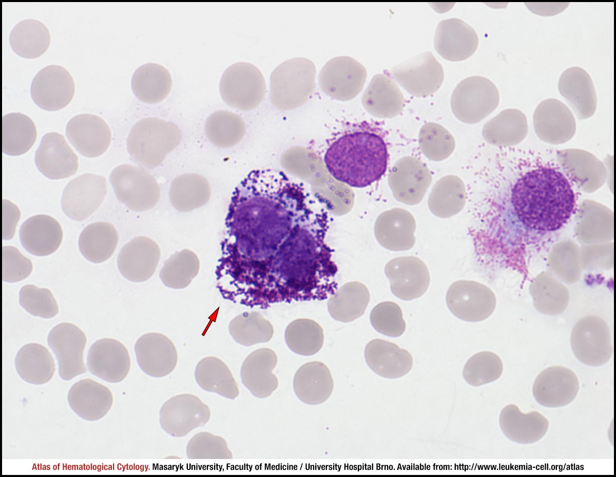 zoom picture
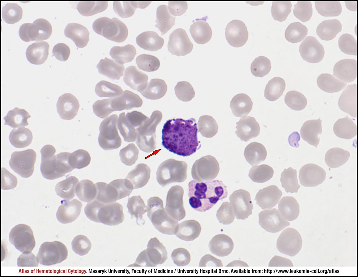 zoom picture
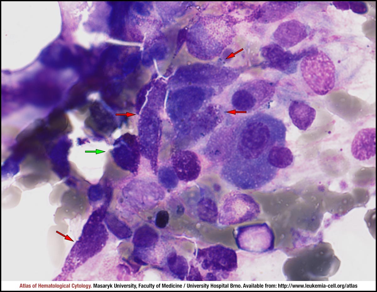 zoom picture
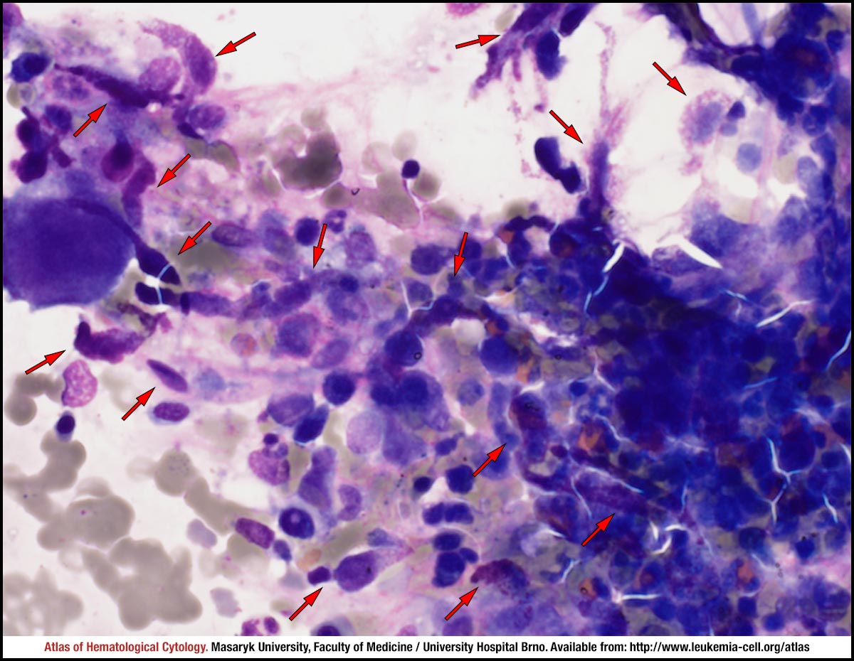 zoom picture