with flowcytometry, cytogenetic and molecular biology findings
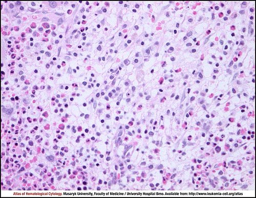
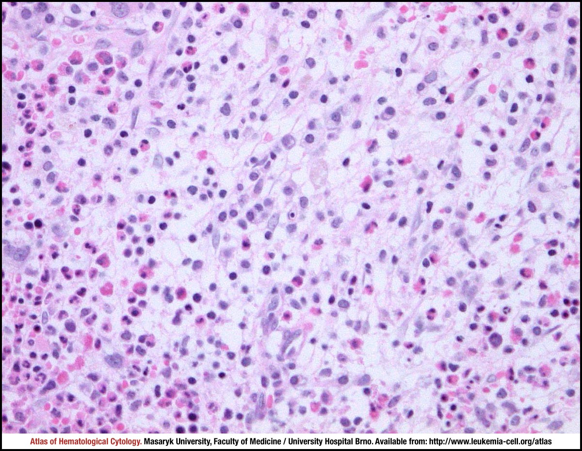
Focal infiltration of bone marrow with fibrosis. The tumour cells with abundant pale cytoplasm are rather inconspicuous in haematoxylin and eosin stain. There is an admixture of small lymphocytes and a large number of eosinophils, which may sometimes dominate and mask the tumour.
Bone marrow trephine biopsy, haematoxylin and eosin stain.
Compared to reactive mast cells rich in metachromatic granules, neoplastic mast cells can be hypogranular or even practically agranular.
Bone marrow trephine biopsy, Giemsa stain.
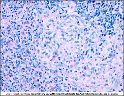
Tumour cells show strong membrane positivity for CD117 (c-KIT). The proof of aggregates of 15 or more mast cells is a major criterion for the diagnosis of systemic mastocytosis.
Bone marrow trephine biopsy, immunohistochemistry.
Atlas of Haematological Cytology [online]. 2016 [cit. 2026-6-25]. Available from WWW: http://www.leukemia-cell.org/atlas.
2026 CELL - Atlas of Haematological Cytology | site map
 zoom picture
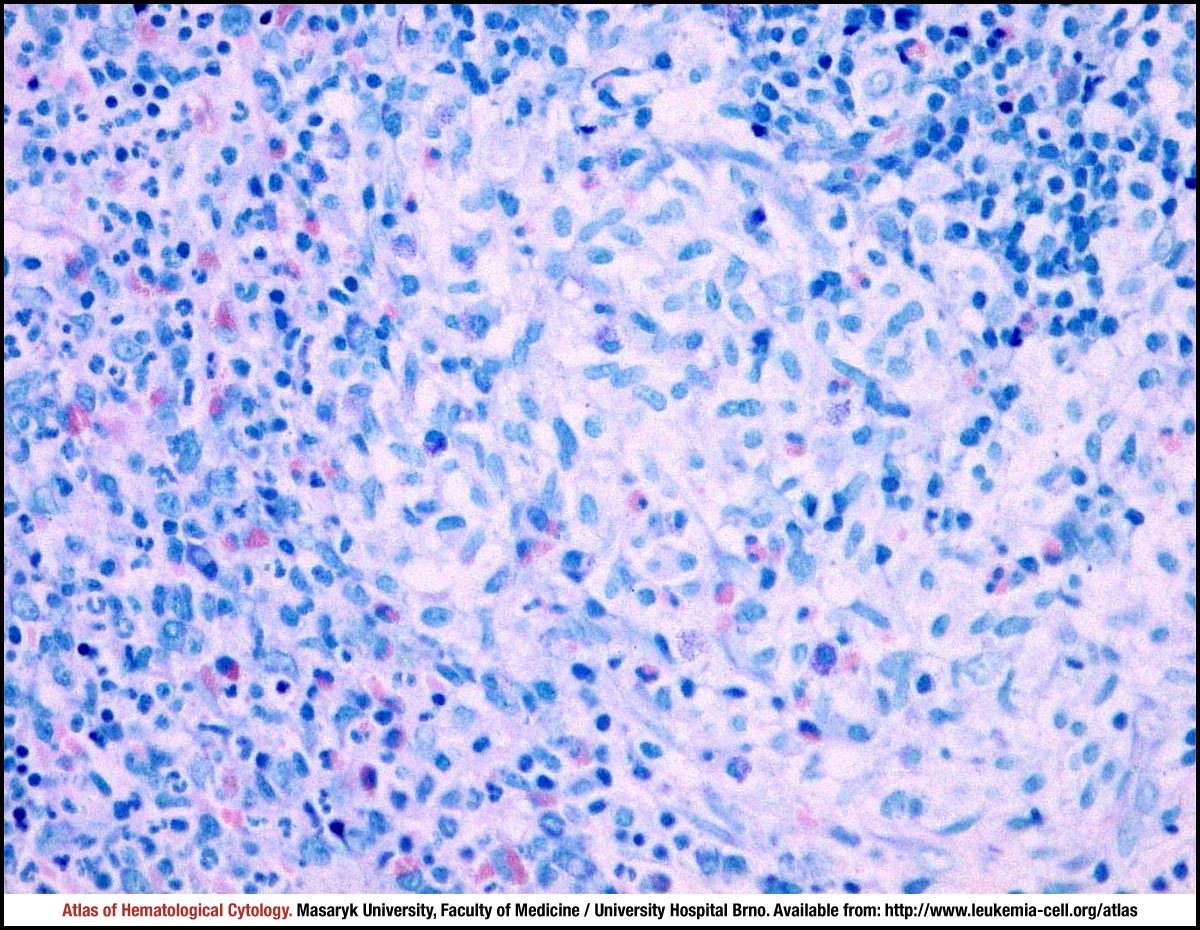 zoom picture
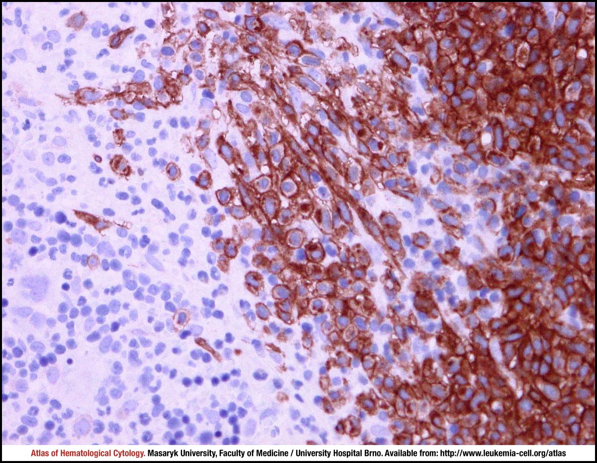 zoom picture