with flowcytometry, cytogenetic and molecular biology findings
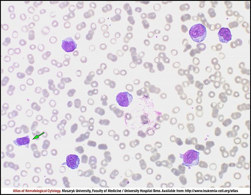
Marked leucocytosis – usually above 100x109/L – is present in the peripheral blood in B-cell prolymphocytic leukaemia. This high leucocyte count is due to the presence of pathological lymphoid cells, the so-called prolymphocytes. Prolymphocytes must account for more than 55% of lymphoid cells in the peripheral blood. Only one lymphocyte (green arrow) is present in this picture.
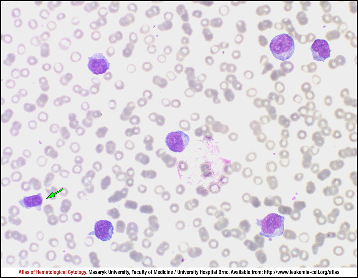
Bone marrow is infiltrated by prolymphocytes, which represent more than 55% of lymphoid cells but also 90% of the all nucleated cells. These prolymphocytes can also occur as smudge cells (green arrow) but their number is significantly lower than in cases of chronic lymphocytic leukaemia.
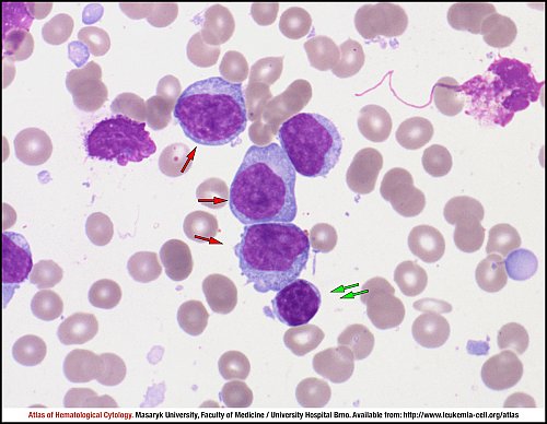
Prolymphocytes (red arrows) are tumour cells in B-cell prolymphocytic leukaemia. The cells are medium to large, twice the size of small lymphocytes (green arrows), with a moderately condensed nuclear chromatin and prominent vesicular nucleoli. The nuclear outline is usually regular and the cytoplasm is weakly basophilic.
Leukaemic cells are less homogenous than those in CLL. They vary in size: larger cells have moderately abundant, weakly basophilic cytoplasm and a round nucleus containing a prominent nucleolus (red arrow), whereas smaller cells tend to have a somewhat higher nuclear-cytoplasmic ratio and their nucleolus is less prominent (green arrow).
Another example of prolymphocytes (red arrows) – tumour cells in B-cell prolyphocytic leukaemia. The bone marrow smear shows cells with relatively plentiful, faintly basophilic cytoplasm, a round nucleus with moderately condensed chromatin, and a single prominent nucleolus.
The analysis of peripheral blood shows a population of B-PLL cells (purple), T lymphocytes (blue), all lymphocytes (green), monocytes (yellow) and granulocytes (red). The population of B-PLL cells expresses CD45 and low SS when compared to normally mature B cells (A); other characteristics involve a positive expression of CD19, CD79b, CD20, a negative expression of CD10 and a clonal expression of the light chain kappa or lambda (B–D); furthermore, histograms show a negative expression of CD23, a positive expression of FMC7 and CD200 and an aberrant positive expression of CD13 (E).
Atlas of Haematological Cytology [online]. 2016 [cit. 2026-6-23]. Available from WWW: http://www.leukemia-cell.org/atlas.
2026 CELL - Atlas of Haematological Cytology | site map
 zoom picture
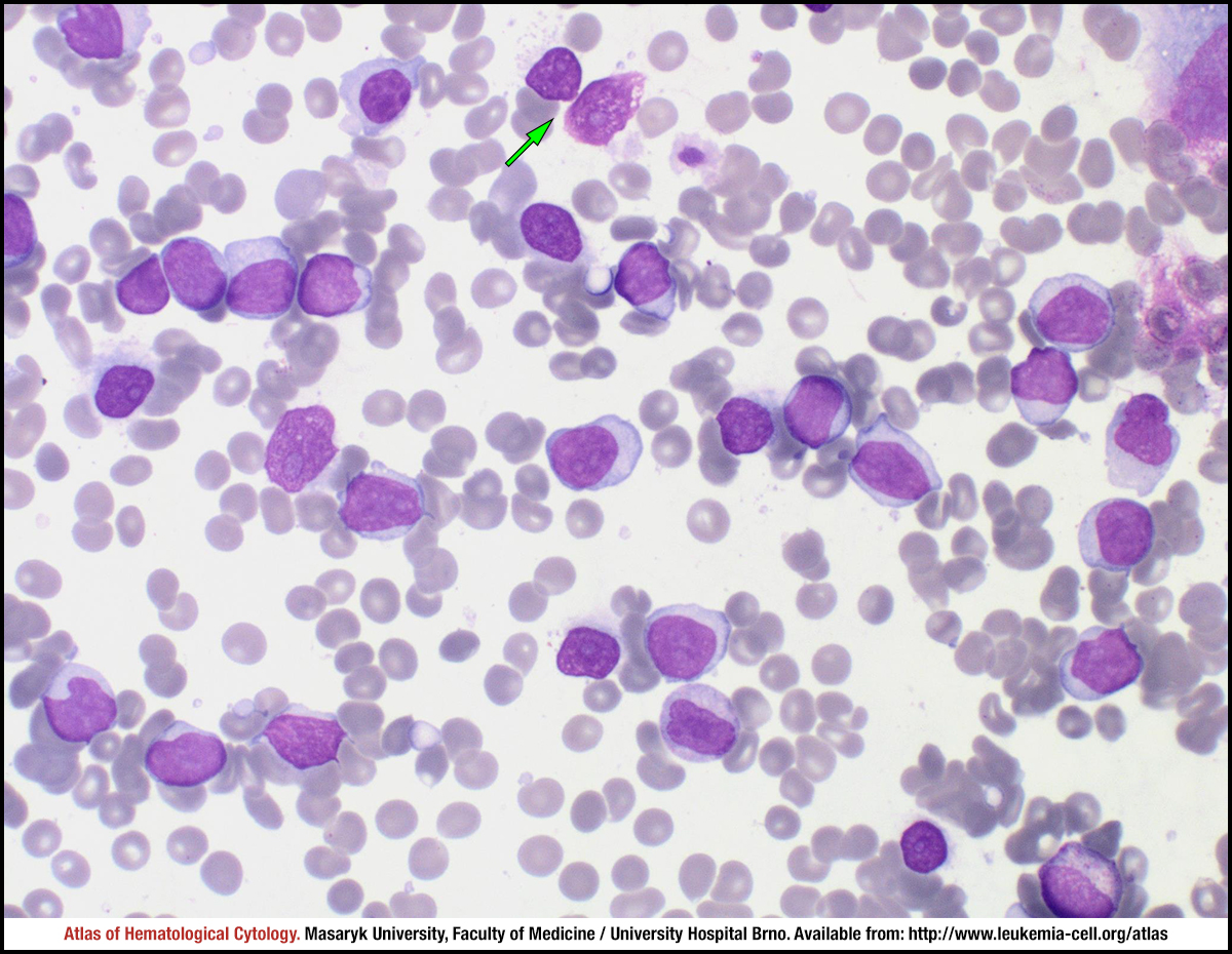 zoom picture
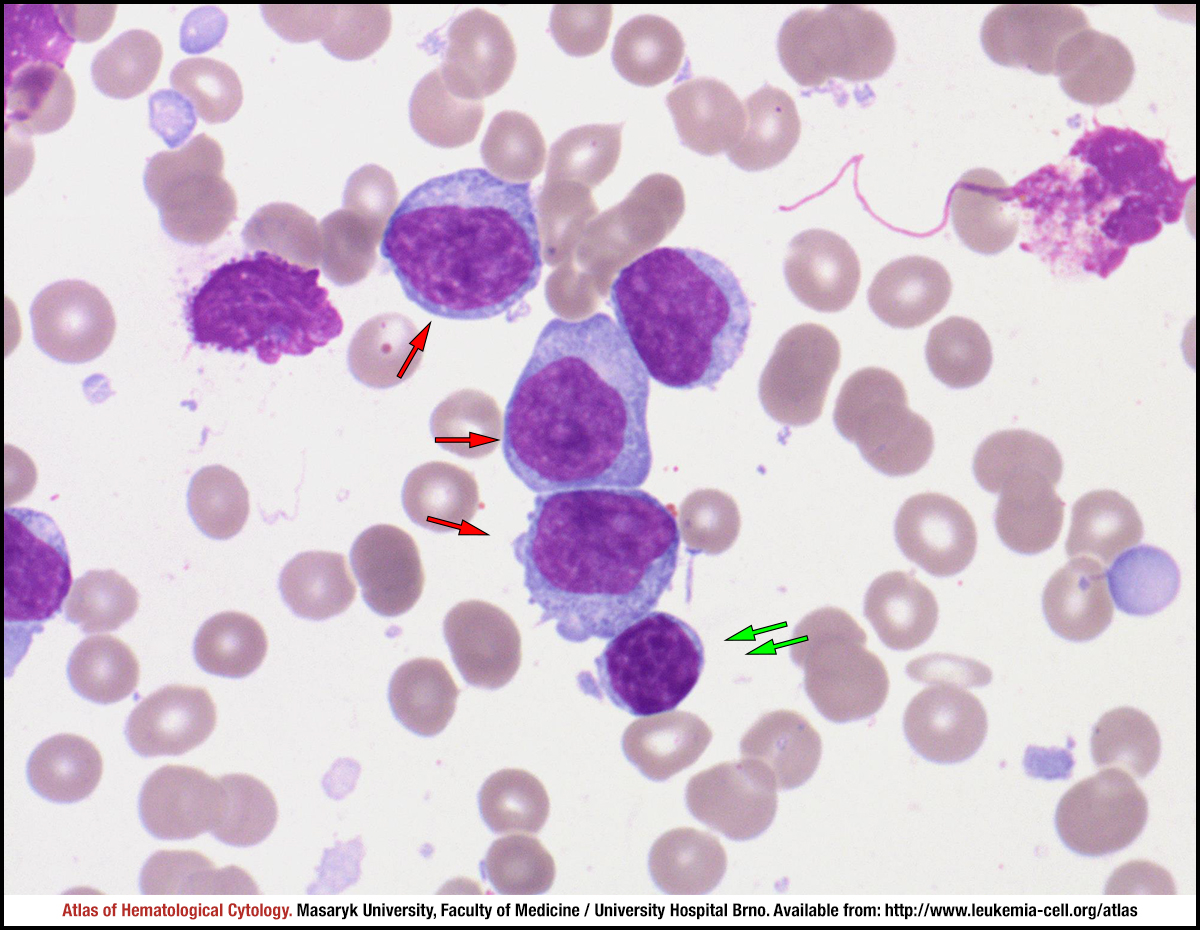 zoom picture
 zoom picture
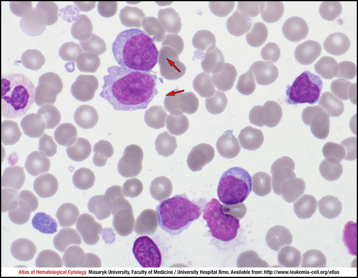 zoom picture
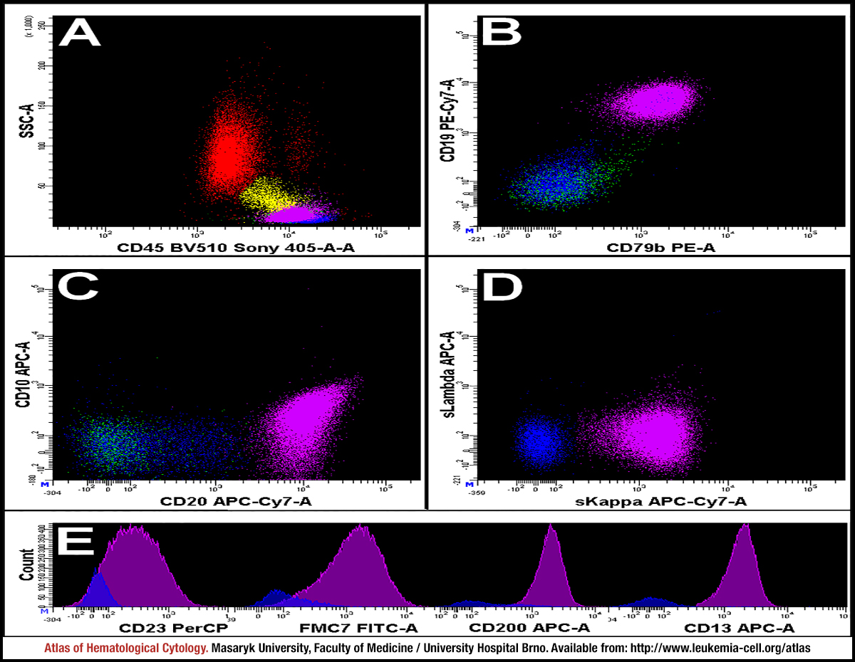 zoom picture