with flowcytometry, cytogenetic and molecular biology findings
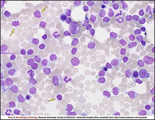
Bone marrow smears shows an infiltration by tumour lymphocytes which is not uniform (as it would be in the classic variant of CLL). In this case of an atypical pleomorphic variant of CLL, more than 10% of tumour cells have a different appearance than typical small lymphocytes, which generally show clumped chromatin and scanty cytoplasm. This morphological variability includes the presence of cells with nucleus indentation and less condensed chromatin; prolymphocytes and a few cells resembling immunoblasts can also occur. The presence of smear cells (yellow arrows) is a typical finding in all cases of CLL.
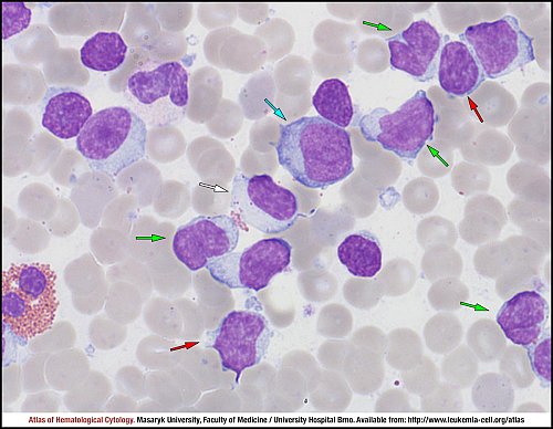
Only several tumour lymphocytes are typical small lymphocytes with clumped chromatin, more or less regular nuclear outlines and scanty cytoplasm (red arrows). Pathological lymphocytes with intended or almost cleft nuclei are present in the smear (green arrows). Tumour cells involve prolymphocytes – larger cells with more abundant cytoplasm and clear nucleoli in nucleus (blue arrow). Not all of the shown lymphocytes can be included to the pathological infiltration: the large granular lymphocyte (white arrow) is a T lymphocyte or natural killer cell.
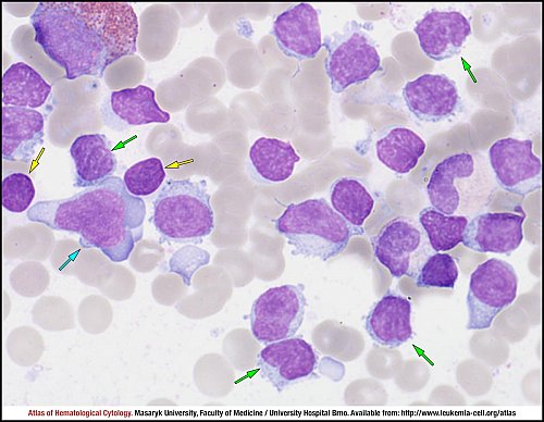
In cases of atypical pleomorphic variants of CLL, some tumour cells can resemble immunoblasts – large cells with a deeply blue cytoplasm and a centrally located nucleus (blue arrow). Many pathological lymphocytes have intended or cleft nuclei (green arrows). Naked smear nuclei, the so-called Gumprecht shadows (yellow arrows), are present as a typical finding in CLL.
Atlas of Haematological Cytology [online]. 2016 [cit. 2024-4-20]. Available from WWW: http://www.leukemia-cell.org/atlas.
2024 CELL - Atlas of Haematological Cytology | site map
 zoom picture
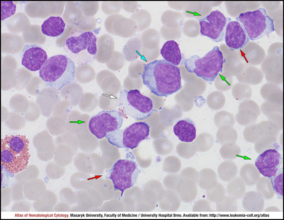 zoom picture
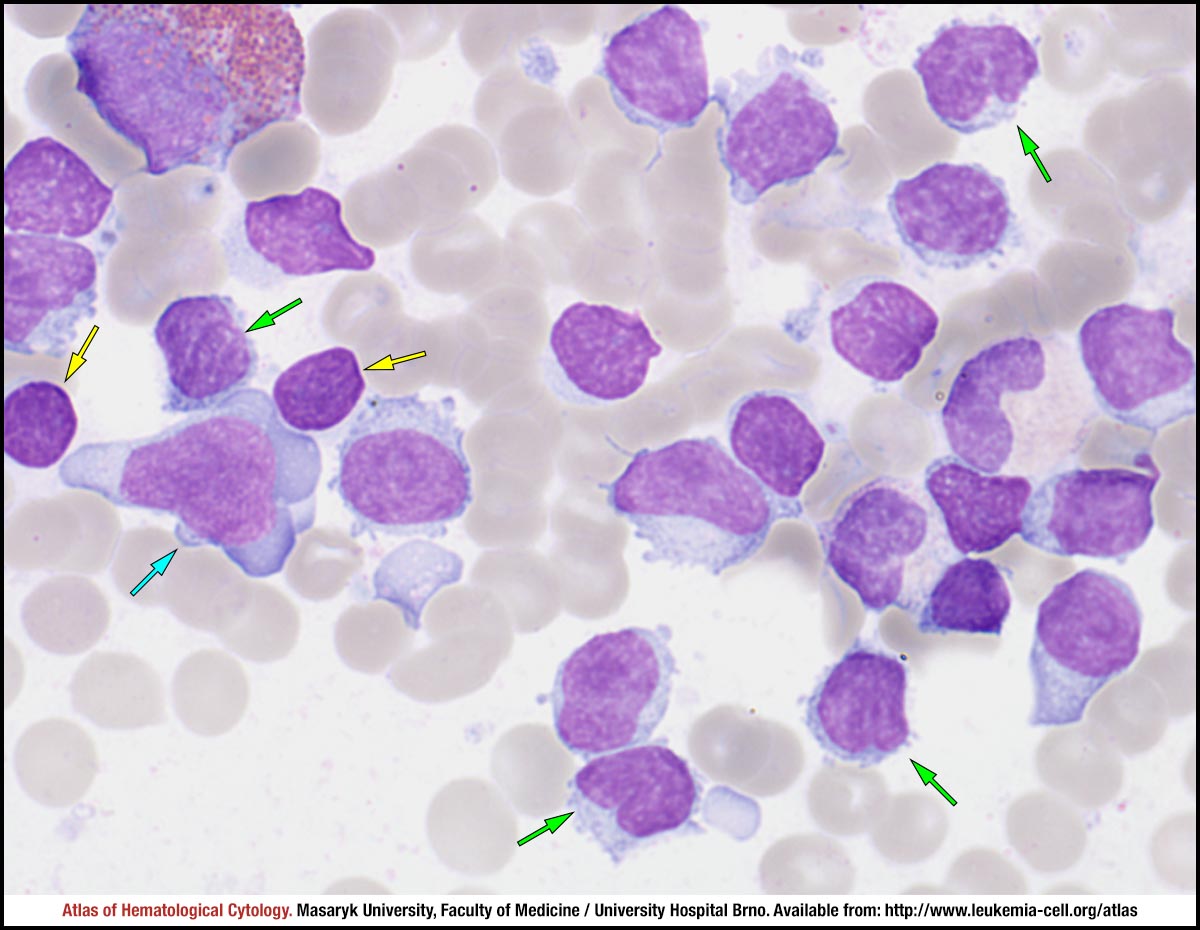 zoom picture