with flowcytometry, cytogenetic and molecular biology findings
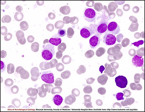
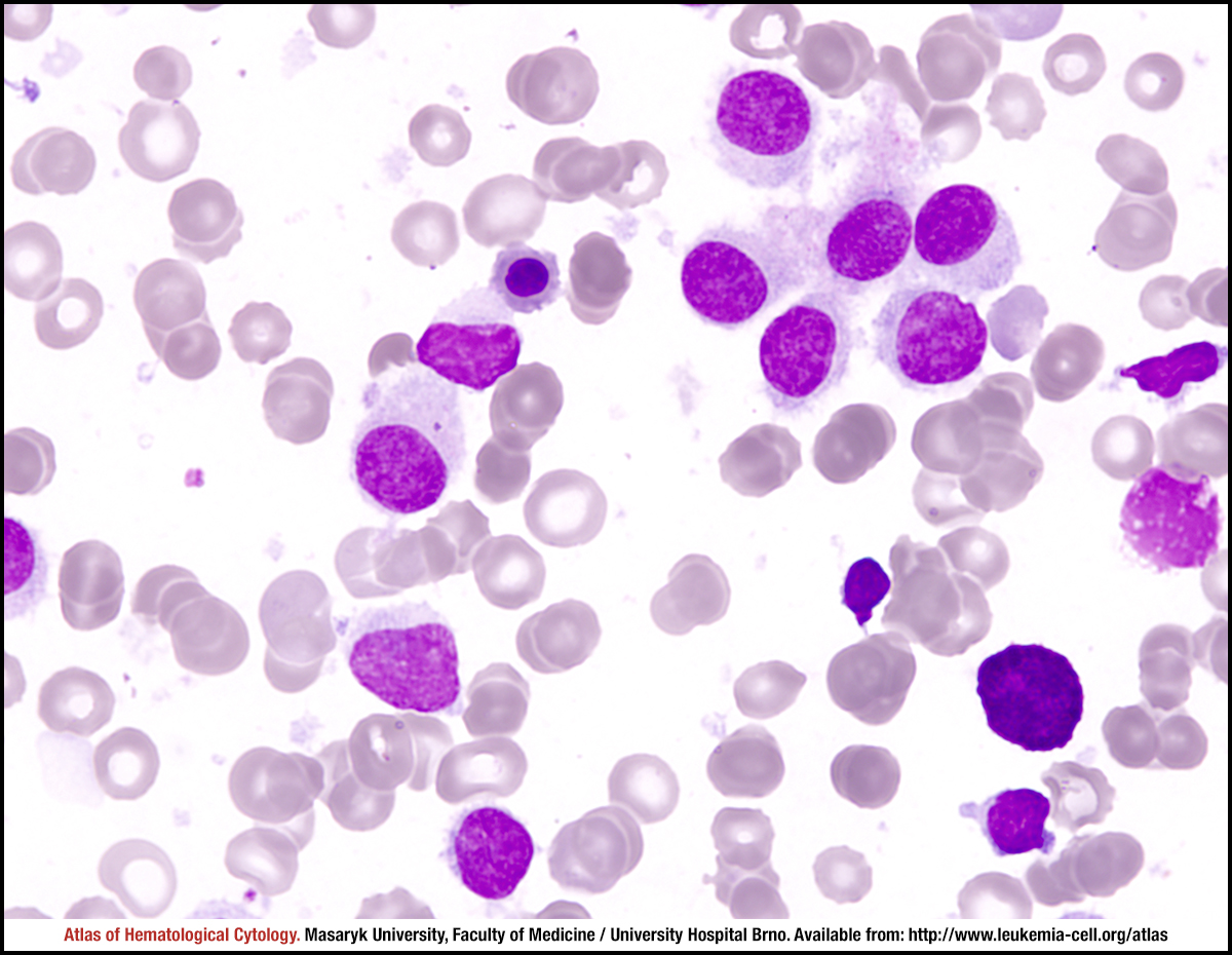
The tumour cells are small to medium-size lymphoid elements with oval, indented (bean-shaped) or occasionally bilobed nucleus with a homogenous spongy, ground-glass chromatin which is slightly less clumped than that in a normal lymphocyte. Nucleoli are typically absent or inconspicuous. The cytoplasm is abundant and pale blue, with circumferential “hairy” projections. The cytoplasm can occasionally contain discrete vacuoles or rod-shaped inclusions.
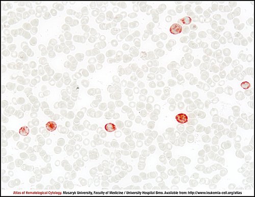
A strong TRAP positivity is characteristic of hairy cell leukaemia. If appropriate air-dried unfixed slide preparations are available (less than 48 hours after preparing), virtually all cases of HCL will contain at least several cells with a strong granular cytoplasmic TRAP positivity (red staining in this case), while weak staining is not diagnostically useful.
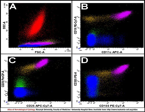
A: HCL cells (purple), all lymphocytes (green), T lymphocytes (blue), normal B lymphocytes (brown), monocytes (yellow) and granulocytes (red). The population of HCL cells has a higher intensity of SS than other lymphocytes.
B, C, D: Characteristic CD19 and CD11c positivity and a higher expression of CD20, CD25, CD103 and CD305 than in normal B lymphocytes.
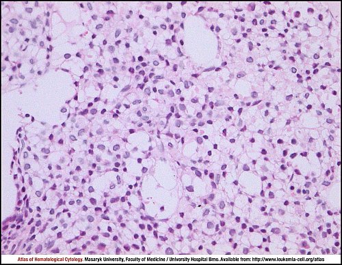
Hairy cell leukaemia. There is a very extensive interstitial to diffuse infiltration of bone marrow.
Bone marrow trephine biopsy, haematoxylin and eosin stain.
Hairy cell leukaemia. The typical loose distribution of small to medium-sized tumour cells due to their abundant pale cytoplasm.
Bone marrow trephine biopsy, haematoxylin and eosin stain.
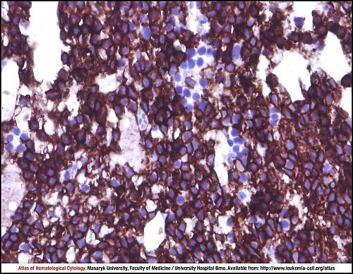
Hairy cell leukaemia. Tumour cells show strong membrane positivity for CD20. There are small clusters of CD20-negative erythropoietic cells.
Bone marrow trephine biopsy, immunohistochemistry.
Atlas of Haematological Cytology [online]. 2016 [cit. 2026-7-11]. Available from WWW: http://www.leukemia-cell.org/atlas.
2026 CELL - Atlas of Haematological Cytology | site map
 zoom picture
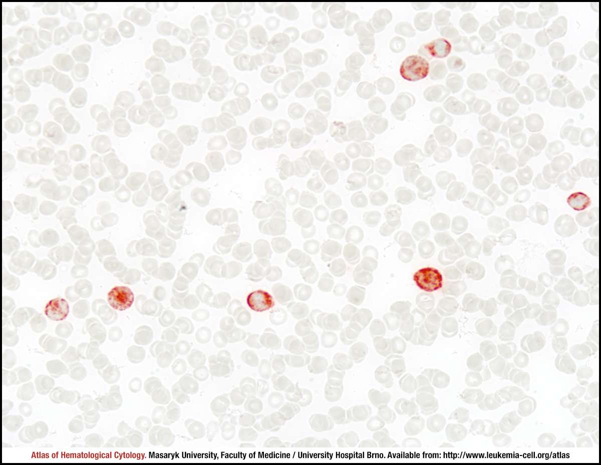 zoom picture
 zoom picture
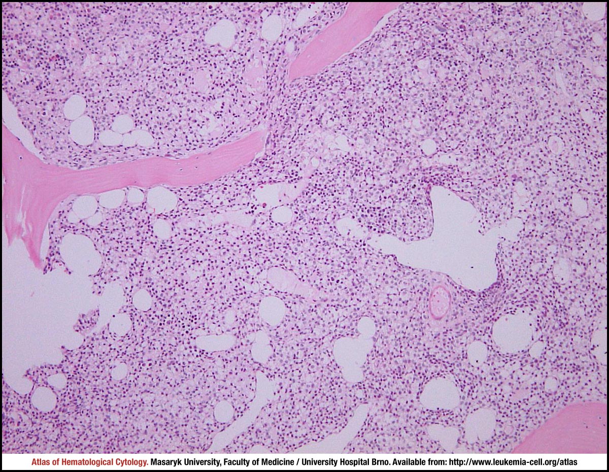 zoom picture
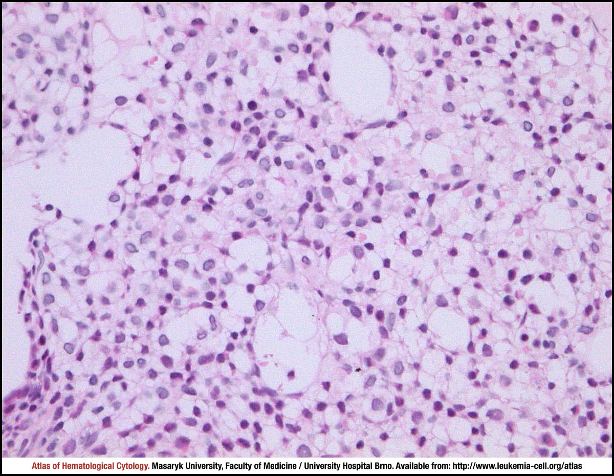 zoom picture
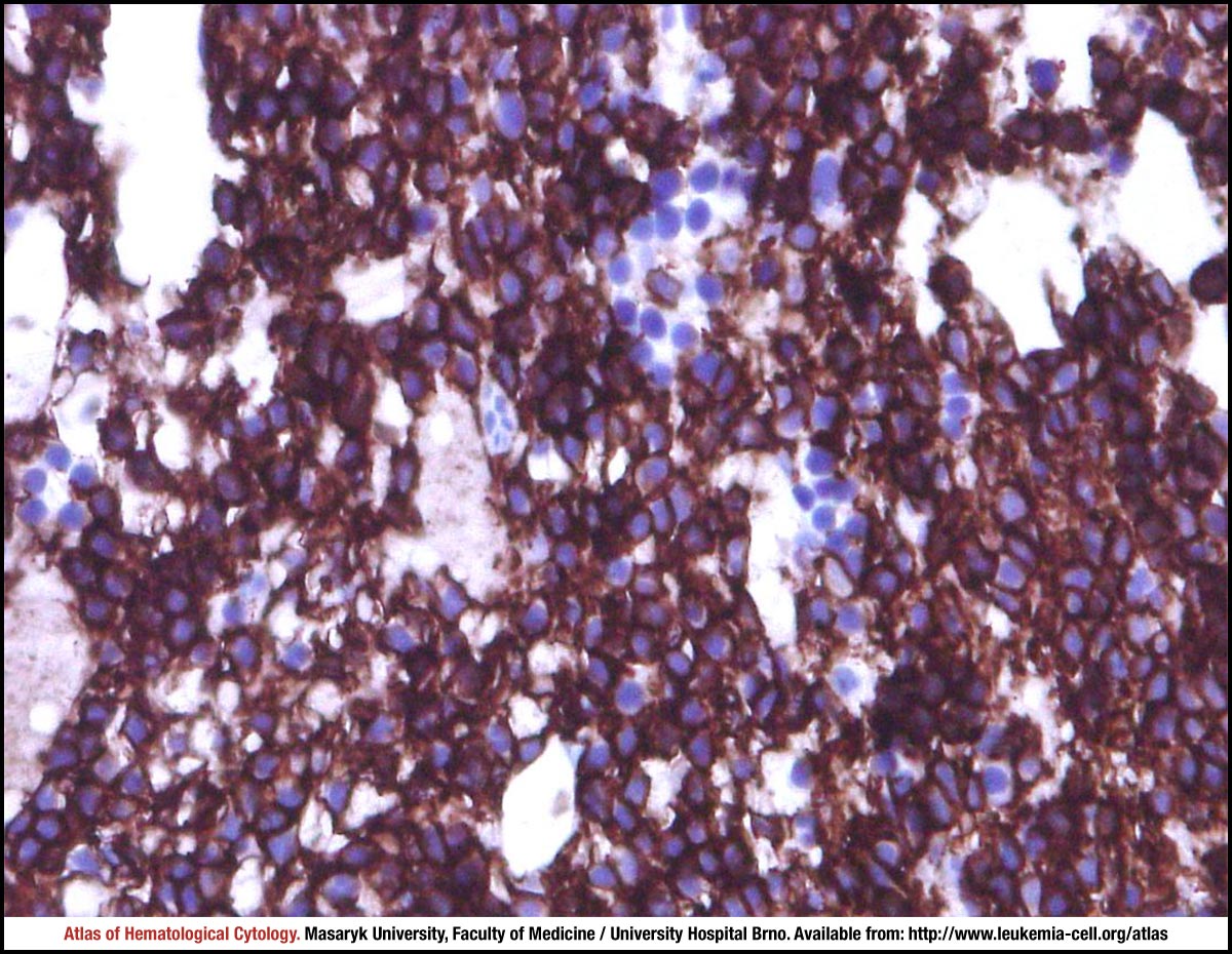 zoom picture