with flowcytometry, cytogenetic and molecular biology findings
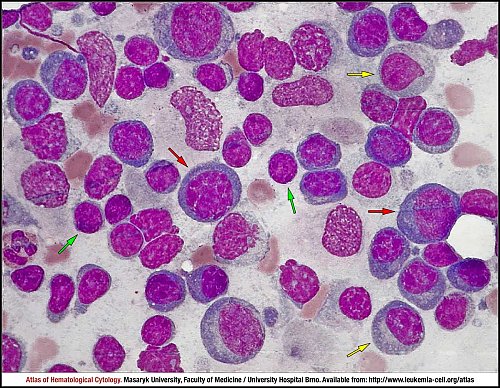
This imprint of lymph node shows an infiltration by lymphoplasmacytic lymphoma. The tumour cells comprise a variable mixture of small lymphocytes with a high nuclear-cytoplasmic ratio, condensed nuclear chromatin and inconspicuous nucleoli (green arrow), medium-sized to large plasmacytoid lymphocytes characterised by an eccentric location of the nucleus, abundant darkly basophilic cytoplasm (yellow arrow) and plasma cells (red arrow).
Atlas of Haematological Cytology [online]. 2016 [cit. 2026-6-23]. Available from WWW: http://www.leukemia-cell.org/atlas.
2026 CELL - Atlas of Haematological Cytology | site map
 zoom picture