with flowcytometry, cytogenetic and molecular biology findings
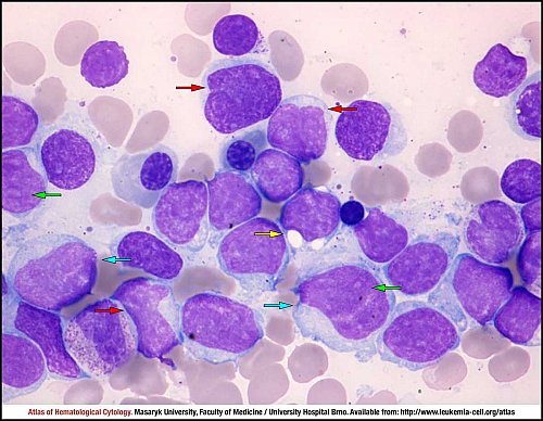
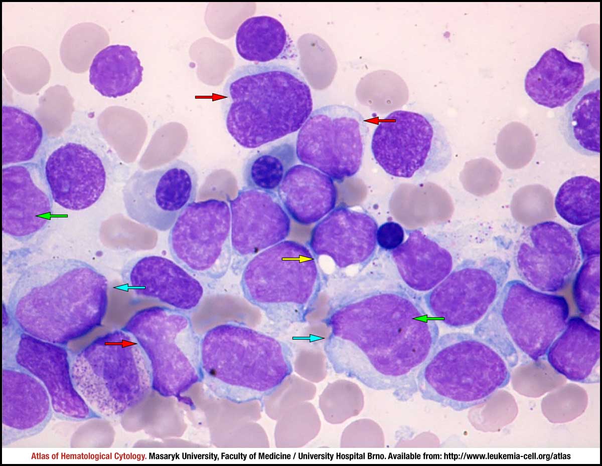
Four morphological variants are distinguished in mantle cell lymphoma. The pleomorphic variant is quite rare and is associated with a more aggressive clinical behaviour of the disease. This bone marrow smear shows a massive infiltration by a mixed pleomorphic population of neoplastic lymphocytes. The size and appearance of tumour cells is not uniform but very variable: large lymphocytes with abundant cytoplasm are present (blue arrows), vacuolisation of the cytoplasm is observed (yellow arrow). The shape of the nucleus is often irregular (red arrows) and some fine nucleoli are visible (green arrows).
The pleomorphic variant with prolymphocytic features is observed in extremely rare cases of mantle cell lymphoma. This peripheral blood smear shows a mixed, highly pleomorphic population of neoplastic lymphocytes. Many tumour cells are medium-sized or large atypical lymphocytes with abundant pale or slightly basophilic cytoplasm and very irregular "floral pattern" shape of the nucleus (blue arrows). Nuclear chromatin is less condensed or fine, prominent large nucleoli are visible (red arrows), which is a typical feature of prolymphocytes.
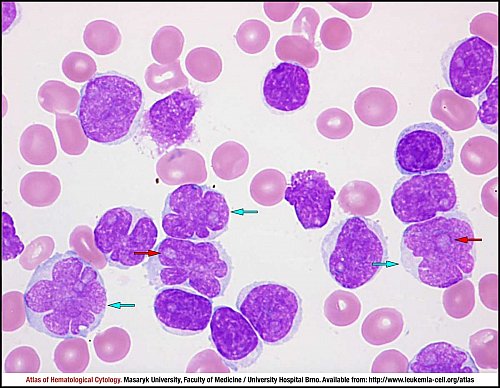
The pleomorphic variant with bilobed nuclei is observed in extremely rare cases of mantle cell lymphoma. This peripheral blood smear shows a predominant population of atypical neoplastic lymphocytes. Tumour cells ale medium-sized lymphocytes with abundant, irregularly shaped basophilic cytoplasm and irregular bilobed nuclei (blue arrows). Nuclear chromatin is less condensed, here with one inconspicuous nucleolus (red arrow).
Atlas of Haematological Cytology [online]. 2016 [cit. 2024-4-25]. Available from WWW: http://www.leukemia-cell.org/atlas.
2024 CELL - Atlas of Haematological Cytology | site map
 zoom picture
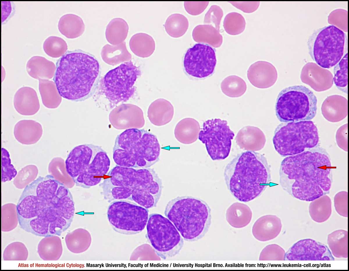 zoom picture
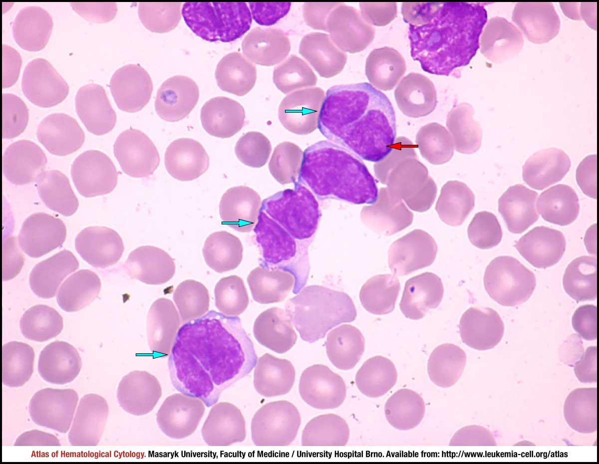 zoom picture