with flowcytometry, cytogenetic and molecular biology findings
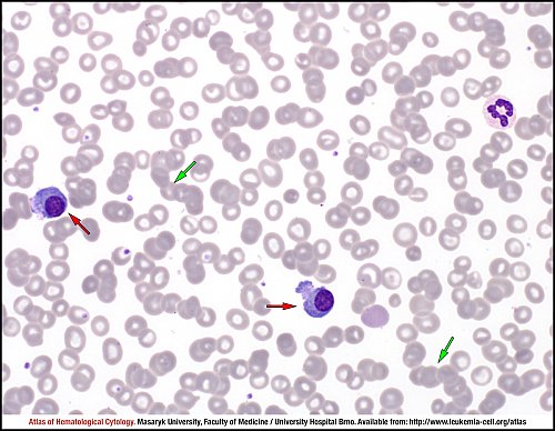
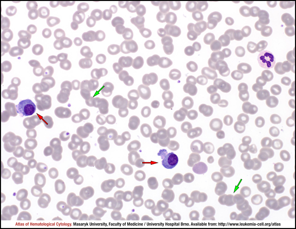
In cases of plasma cell leukaemia, the number of clonal plasma cell (red arrows) in peripheral blood exceeds 2x10⁹/L or is at least 20% of the leucocyte differential count. The smear also shows the so-called “rouleaux” formation of erythrocytes (green arrows).
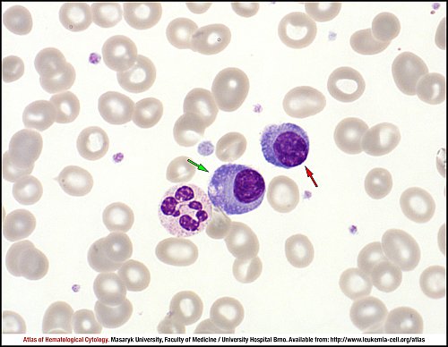
A tumour cell in plasma cell leukaemia may be morphologically fairly normal (green arrow), showing the eccentric nucleus, clumped chromatin and Golgi zone, which are all typical features of a normal plasma cell. Cytoplasmic vacuolisation might occur in both cases – in normal plasma cells and in myeloma cells. A smaller tumour cell with relatively little cytoplasm, relatively higher nuclear-cytoplasmic ratio and without a clear Golgi zone (red arrow) is a more frequent finding in plasma cell leukaemia in peripheral blood.
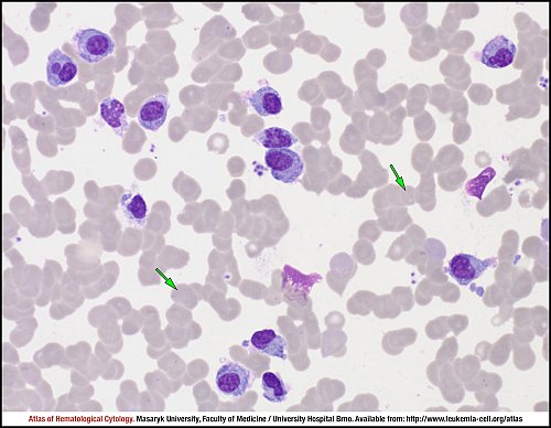
This picture shows numerous tumour cells found in bone marrow in cases of plasma cell leukaemia. The cells vary mainly in the shape of their nuclei and in maturity. The “rouleaux” formation of erythrocytes is clearly visible (green arrows).
Spherical inclusions containing weakly basophilic material, the so-called Russell bodies (green arrow), are present in the cytoplasm of myeloma cells. Tumour cells can also have other cytoplasmic abnormalities, such as cytoplasmic eosinophilia (red arrows).
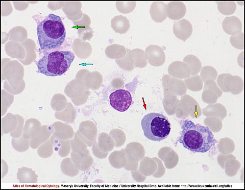
Different morphological types of tumour cells can be present in bone marrow in cases of plasma cell leukaemia. A myeloma cell can be rather mature, resembling a normal plasma cell (yellow arrow); the only visible difference is a slightly more mature chromatin structure. Another tumour cell (green arrow) is morphologically less mature, with a clear nucleolus in the nucleus; such cells are often referred to as proplasmocytes. Yet another tumour cell (cyan arrow) with a higher nuclear-cytoplasmic ratio, multiple nucleoli in the nucleus and a finer chromatin structure would be probably classified as a plasmablast. Erythroid precursors must be distinguished from tumour cells; an intermediate (polychromatic) erythroblast with a slightly megaloblastoid structure of the nucleus (red arrow) is shown in the picture.
Atlas of Haematological Cytology [online]. 2016 [cit. 2024-4-19]. Available from WWW: http://www.leukemia-cell.org/atlas.
2024 CELL - Atlas of Haematological Cytology | site map
 zoom picture
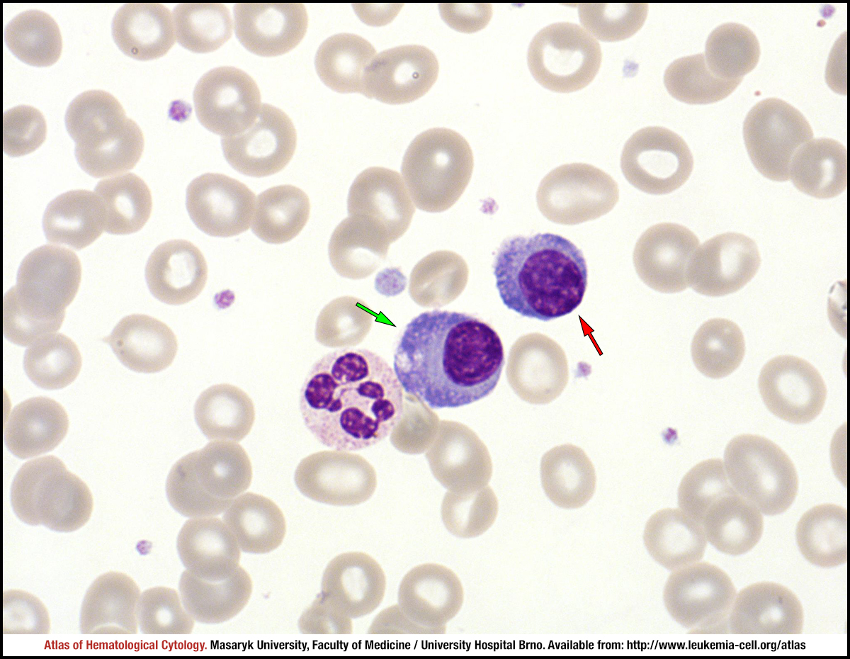 zoom picture
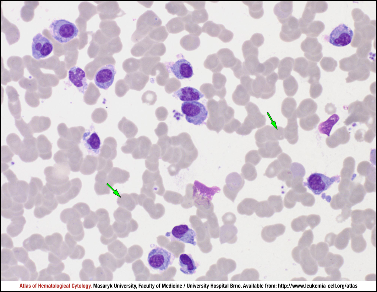 zoom picture
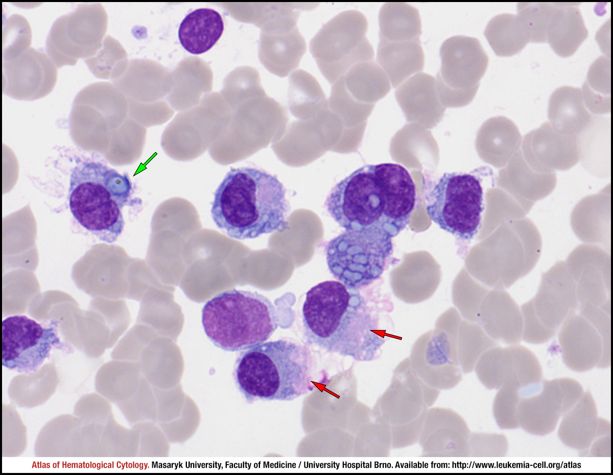 zoom picture
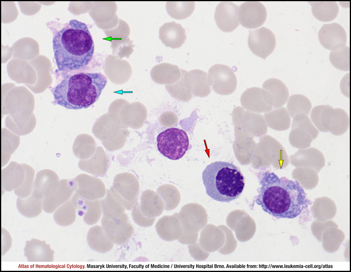 zoom picture