with flowcytometry, cytogenetic and molecular biology findings
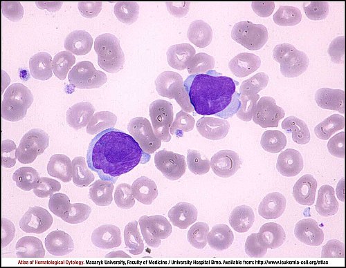
Sézary syndrome is a rare aggressive subtype of cutaneous T-cell lymphoma. It is closely related to mycosis fungoides and is presented with erythroderma, generalised lymphadenopathy and circulating malignant T cells (Sézary cells). This peripheral blood smear shows two typical Sézary cells. The cells are medium-sized to large with abundant, irregularly shaped and basophilic cytoplasm. The shape of the nucleus is very irregular, hyperconvoluted with a cerebriform pattern. Nuclear chromatin is condensed, nucleoli are not visible. Cerebriform appearance of the nucleus is a typical finding in Sézary syndrome.
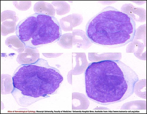
Four cutouts of a peripheral blood smear picture show the typical cytomorphology of Sézary cells. The cells are medium-sized to large with abundant, irregularly shaped and basophilic cytoplasm. The shape of the nucleus is very irregular, hyperconvoluted with a cerebriform pattern. Nuclear chromatin is condensed, nucleoli are not visible. Cerebriform appearance of the nucleus is a typical finding in Sézary syndrome.
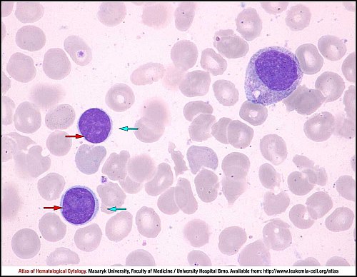
Bone marrow involvement is rare in T-cell lymphomas but may be present in Sézary syndrome. This hypocellular bone marrow smear shows two neoplastic T cells, the so-called Lutzner cells (blue arrows). Unlike classical Sézary cells, these tumour lymphocytes are small, with a high nuclear-cytoplasmic ratio and a spare amount of slightly basophilic cytoplasm. Rounded nuclei have condensed chromatin, nucleoli are not present and inconspicuous indentations of nuclear margin (red arrows) are visible in careful observation.
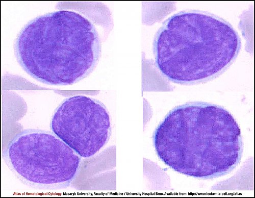
Four cutouts of peripheral blood smear picture show the typical cytomorphology of Lutzner cells. The tumour lymphocytes are small, with a high nuclear-cytoplasmic ratio and a spare amount of slightly basophilic cytoplasm. Rounded or oval nuclei are characterised by condensed chromatin, nucleoli are not present and irregular indentations of nucleus are visible in careful observation.
Atlas of Haematological Cytology [online]. 2016 [cit. 2026-6-25]. Available from WWW: http://www.leukemia-cell.org/atlas.
2026 CELL - Atlas of Haematological Cytology | site map
 zoom picture
 zoom picture
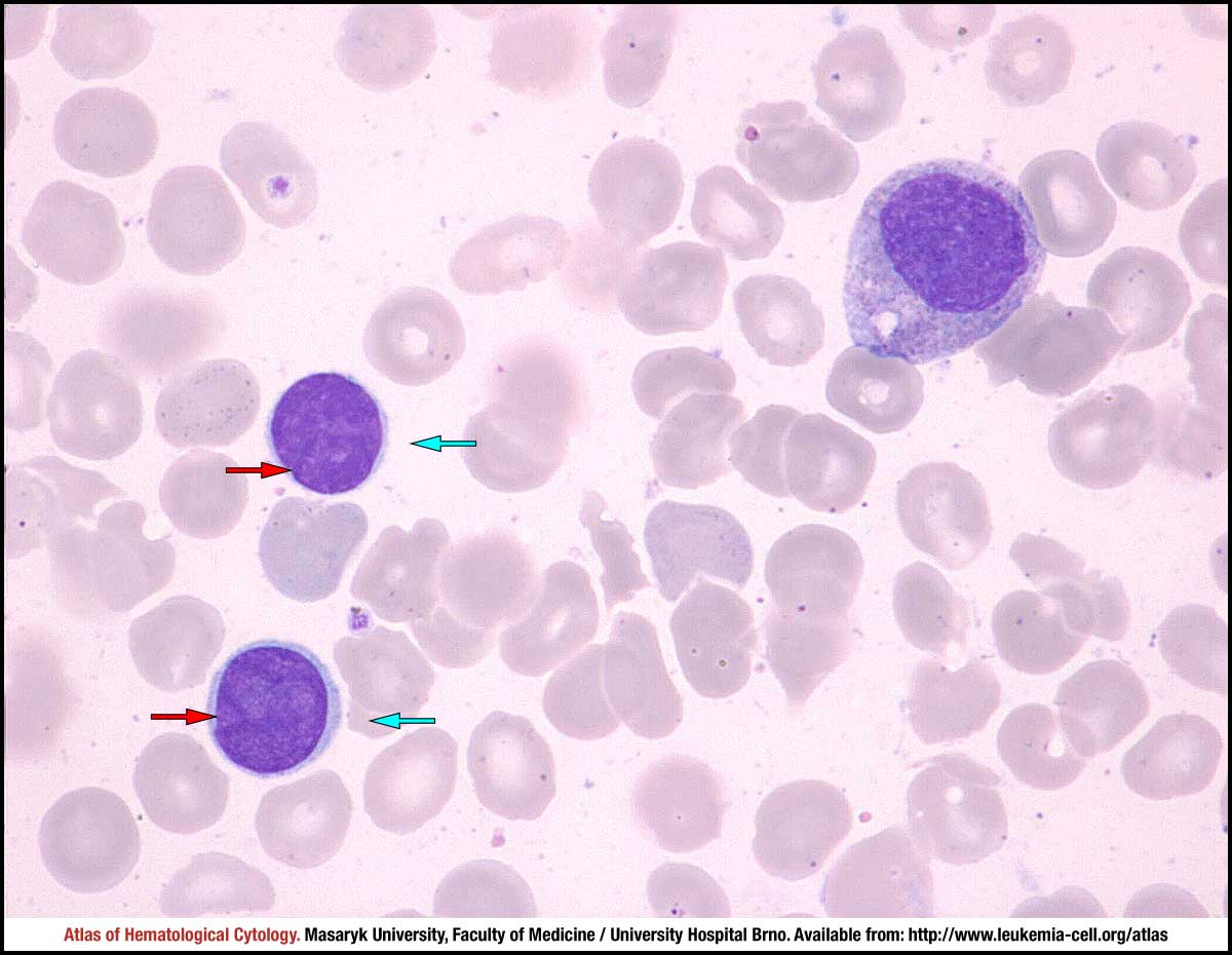 zoom picture
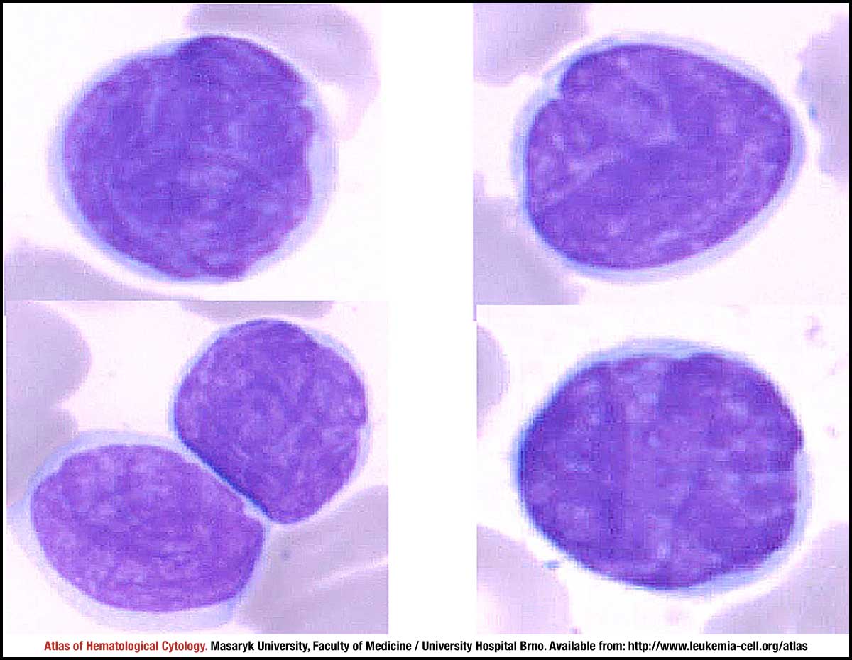 zoom picture