with flowcytometry, cytogenetic and molecular biology findings
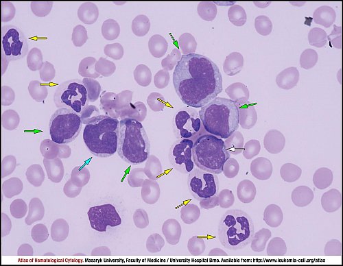
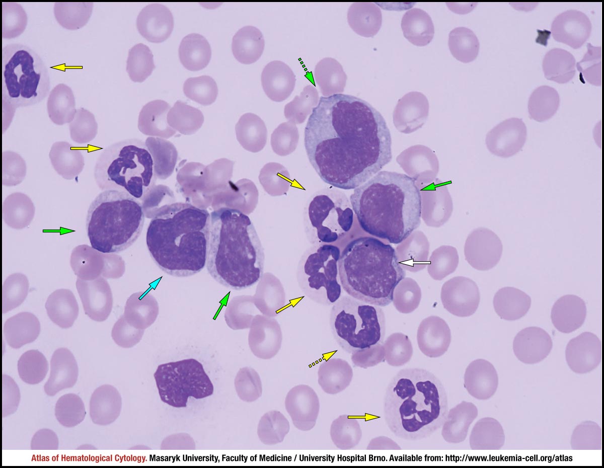
Solid green arrows mark three atypical monocytes: they are slightly larger and show a decreased lobularity of the nucleus. A large promonocyte with mild vacuolation of cytoplasm is marked by a dotted green arrow. Hypogranular/agranular segmented neutrophils are marked by solid yellow arrows and a neutrophilic band cell is marked by a dotted yellow arrow. The white arrow marks a myelocyte with a lower amount of cytoplasm, which shows a decreased granularity. The cyan arrow marks a megaloblastoid metamyelocyte with hypogranular cytoplasm.
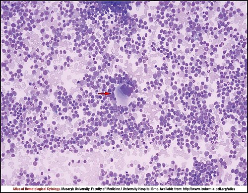
Marrow aspirate smear with an increased cellularity and a predominant population of myelomonocytic cells. In the centre of the image, the red arrow marks a megakaryocyte larger in size, with a hyperlobulated nucleus and hypogranular cytoplasm.
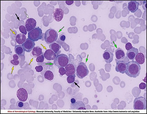
Pathological monocytes are marked by green arrows: a monoblast (solid arrow), a monocyte (dashed arrow) and two promonocytes (dotted arrows). The image also shows hypogranular neutrophils in different stages of maturation (yellow arrows), two lymphocytes (black arrows) and a larger atypical eosinophil containing basophilic progranules (white arrow). Unmarked cells correspond to erythroblasts, largely with megaloblastoid features.
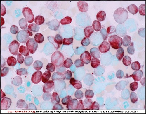
Alpha-naphthyl butyrate esterase (ANBE) staining: monocytes (the majority of cells shown in the image) stain with various intensity, the reaction product is brown and diffuse. Other cells shown in the image (erythroblasts and neutrophils) do not stain in this method.
Atlas of Haematological Cytology [online]. 2016 [cit. 2025-7-14]. Available from WWW: http://www.leukemia-cell.org/atlas.
2025 CELL - Atlas of Haematological Cytology | site map
 zoom picture
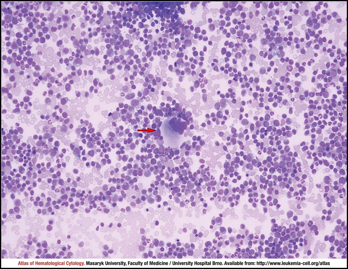 zoom picture
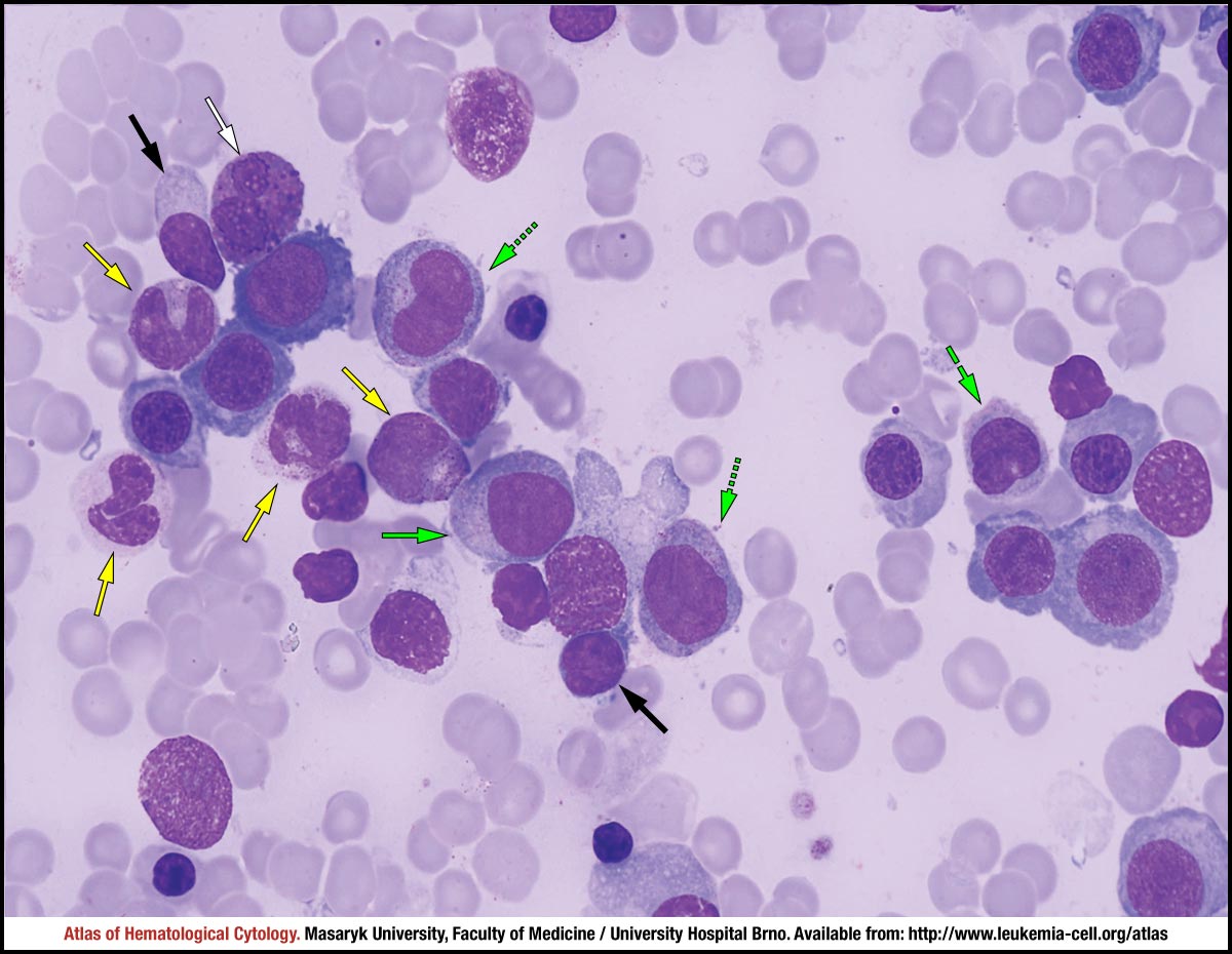 zoom picture
 zoom picture