with flowcytometry, cytogenetic and molecular biology findings
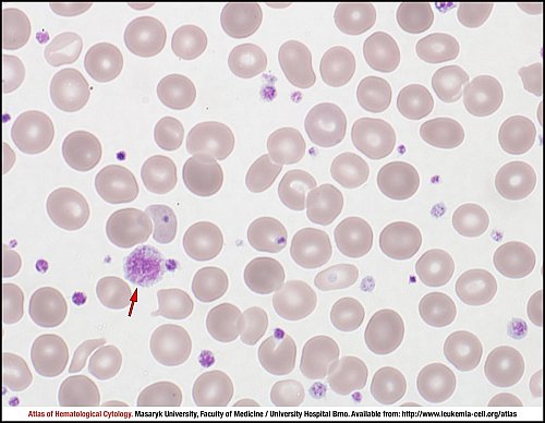
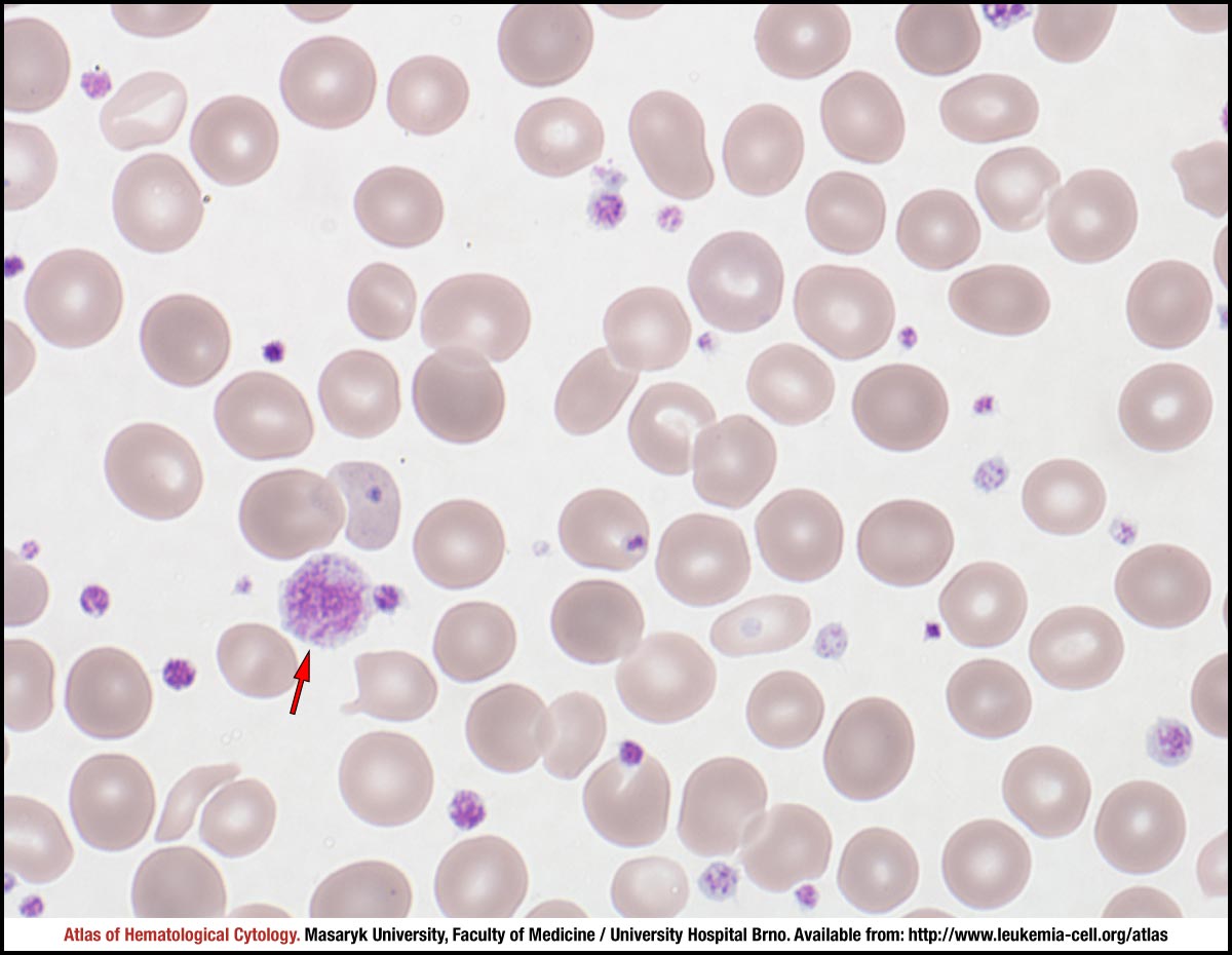
The peripheral blood smear shows marked thrombocytosis with occasional macrothrombocytes (red arrow). Diagnostic criteria of MDS/MPN-RS-T involve a persistently elevated platelet count (≥ 450 × 109/L) as well as anaemia in the peripheral blood.
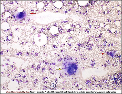
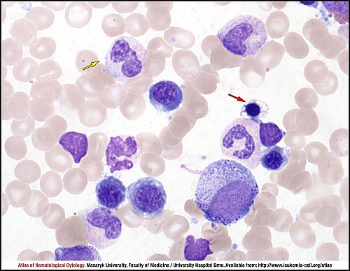
The bone marrow smear is slightly hypercellular. It shows three megakaryocytes in the visible field, which is a sign of rather increased megakaryopoiesis. MDS/MPN-RS-T is usually characterised by megakaryocytes that are rather large and mature (red arrows) or almost giant (yellow arrow). The morphological appearance of these elements resembles megakaryocytes that are seen in other myeloproliferative diseases such as essential thrombocythaemia or primary myelofibrosis.
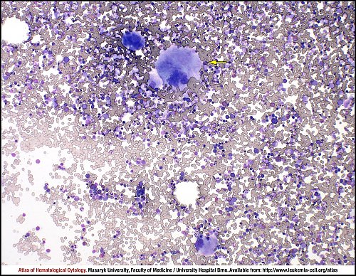
Bone marrow is hypercellular in this region of bone marrow smear and a number of megakaryocytes are visibly increased (four elements in total). One of them (yellow arrow) is giant and resembles a megakaryocyte usually seen in essential thrombocythaemia.
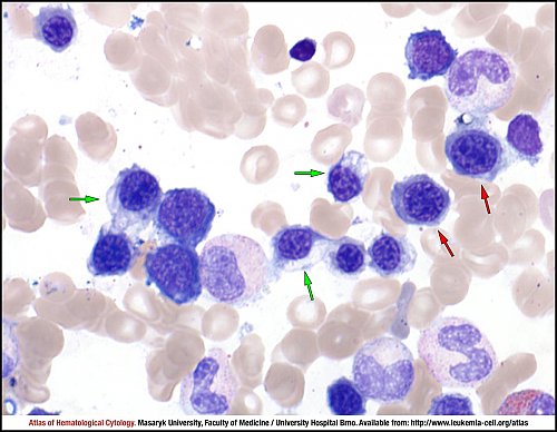
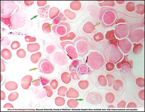
The bone marrow smear shows increased erythropoiesis due to an ineffective erythroid proliferation, with megaloblastoid (red arrows) and/or other dyserythropoietic features, such as erythroid precursors with poorly haemoglobinised or vacuolated cytoplasm (green arrows).
Presence of erythroid precursors with vacuolated cytoplasm and basophilic stippling (red arrow) is the most typical morphological finding in MGG staining. Dysplastic features can also occur in granulopoiesis; an abnormal nuclear clumping is present in this case (yellow arrow). Single lineage dysplasia (erythroid-lineage) or, more rarely, multilineage dysplasia is characteristic for the diagnosis of MDS/MPN-RS-T.
The diagnosis is of MDS/MPN-RS-T is ruled out by an increased proportion of blasts in WBC differential in the peripheral blood (> 1%) and/or by an increased proportion of blasts in all nucleated cells in the bone marrow differential count (> 5%).
In addition to typical dyserythropoietic features such as late erythroid precursors with vacuolated cytoplasm and basophilic stippling (red arrow), other morphological changes can also be present. The green arrow points to an abnormal mature erythropoietic precursor with abundant vacuolated/poorly haemoglobinised cytoplasm and an abnormal nuclear-cytoplasmic ratio.
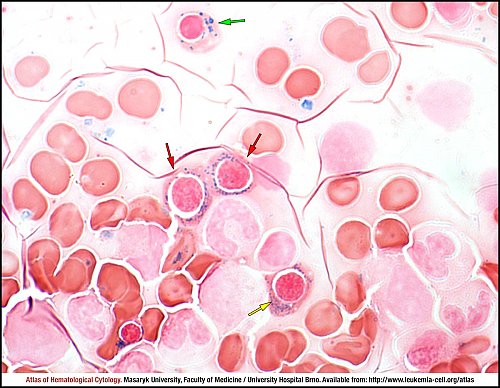
Although dyserythropoiesis is the most striking feature in the cytomorphological examination in May-Grünwald-Giemsa staining, other dysplastic changes in various morphological lineages can also occur. Dyserythropoietic changes shown in this particular part of bone marrow smear involve one megaloblastoid erythroid precursor (green arrow), abnormal clumping of nuclear chromatin (yellow arrow) and strangulated nuclei (red arrow) in a late erythroblast.
Perls’ or Prussian blue reaction displays iron in different compartments of bone marrow cells. This bone marrow smear shows iron in siderocytes (green arrows), in sideroblasts (yellow arrows) and in ring sideroblasts (red arrows). The presence of ring sideroblasts accounting for ≥ 15% of erythroblasts belongs to diagnostic criteria. A ring sideroblast is defined as an erythroid precursor with more than five siderotic granules present in a perinuclear position, surrounding the nucleus or encompassing at least one third of the nuclear circumference.
The bone marrow smear shows another example of ring sideroblasts (red arrows) occurring in MDS/MPN-RS-T, which are hallmarks of this diagnosis. Other abnormal sideroblasts can also occur, such as cells with siderotic granules that are either present in large numbers (yellow arrow) or are abnormally large (green arrow).
Atlas of Haematological Cytology [online]. 2016 [cit. 2026-7-11]. Available from WWW: http://www.leukemia-cell.org/atlas.
2026 CELL - Atlas of Haematological Cytology | site map
 zoom picture
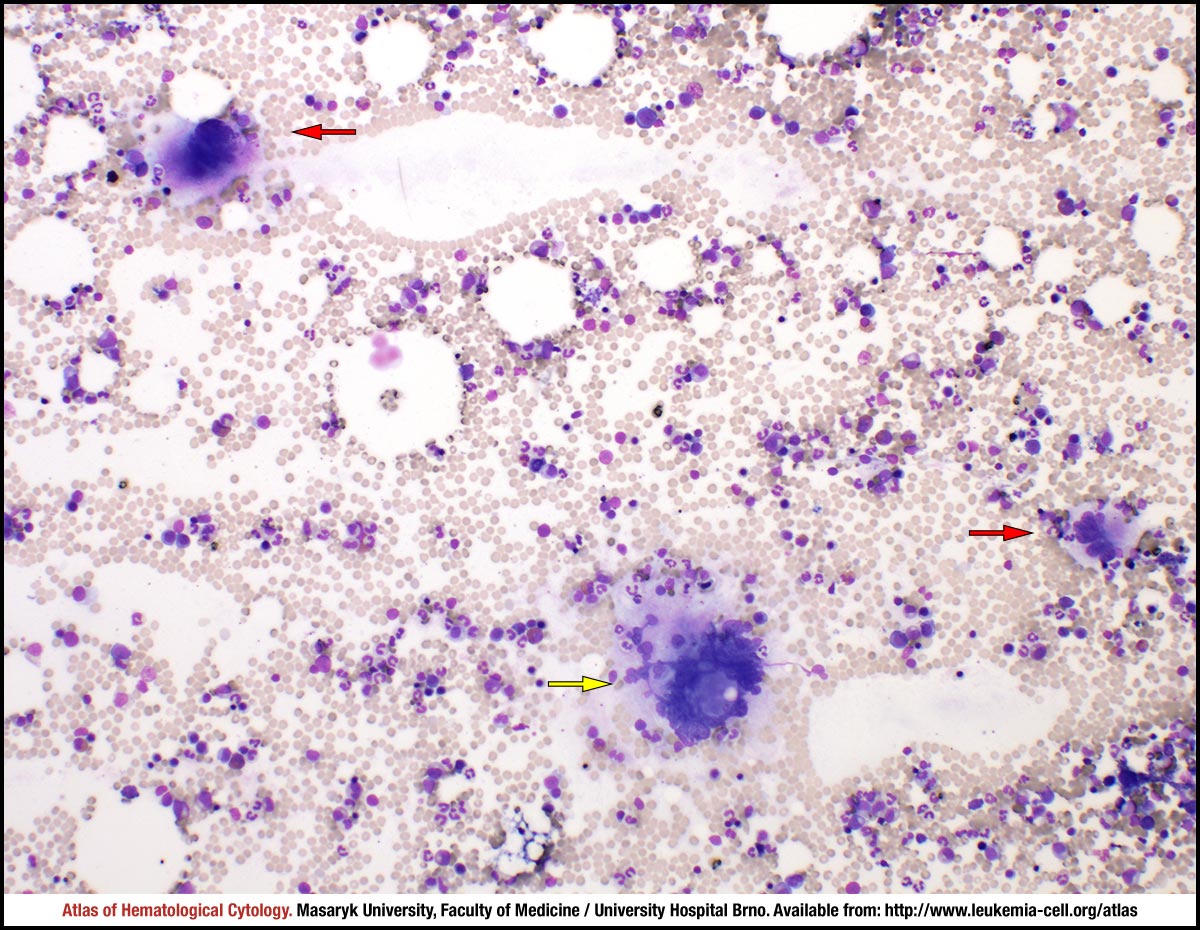 zoom picture
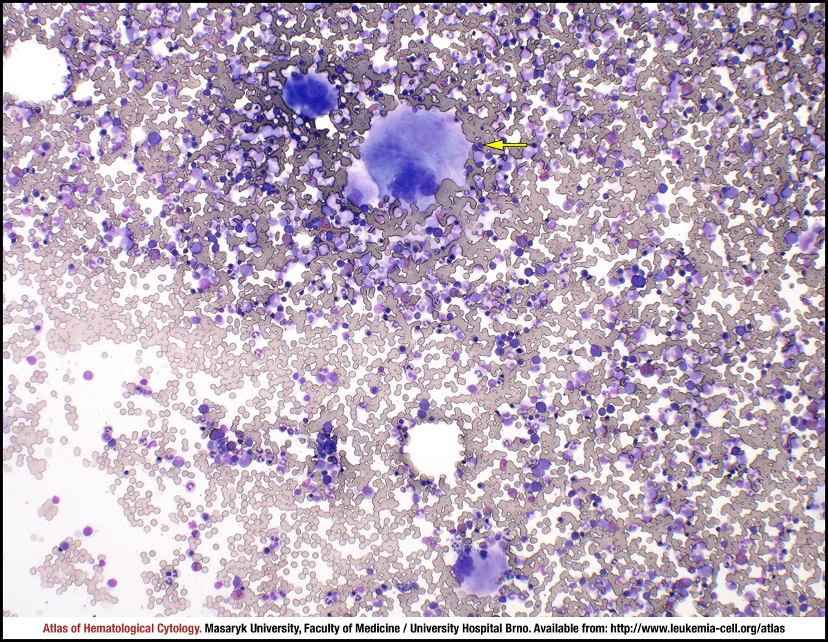 zoom picture
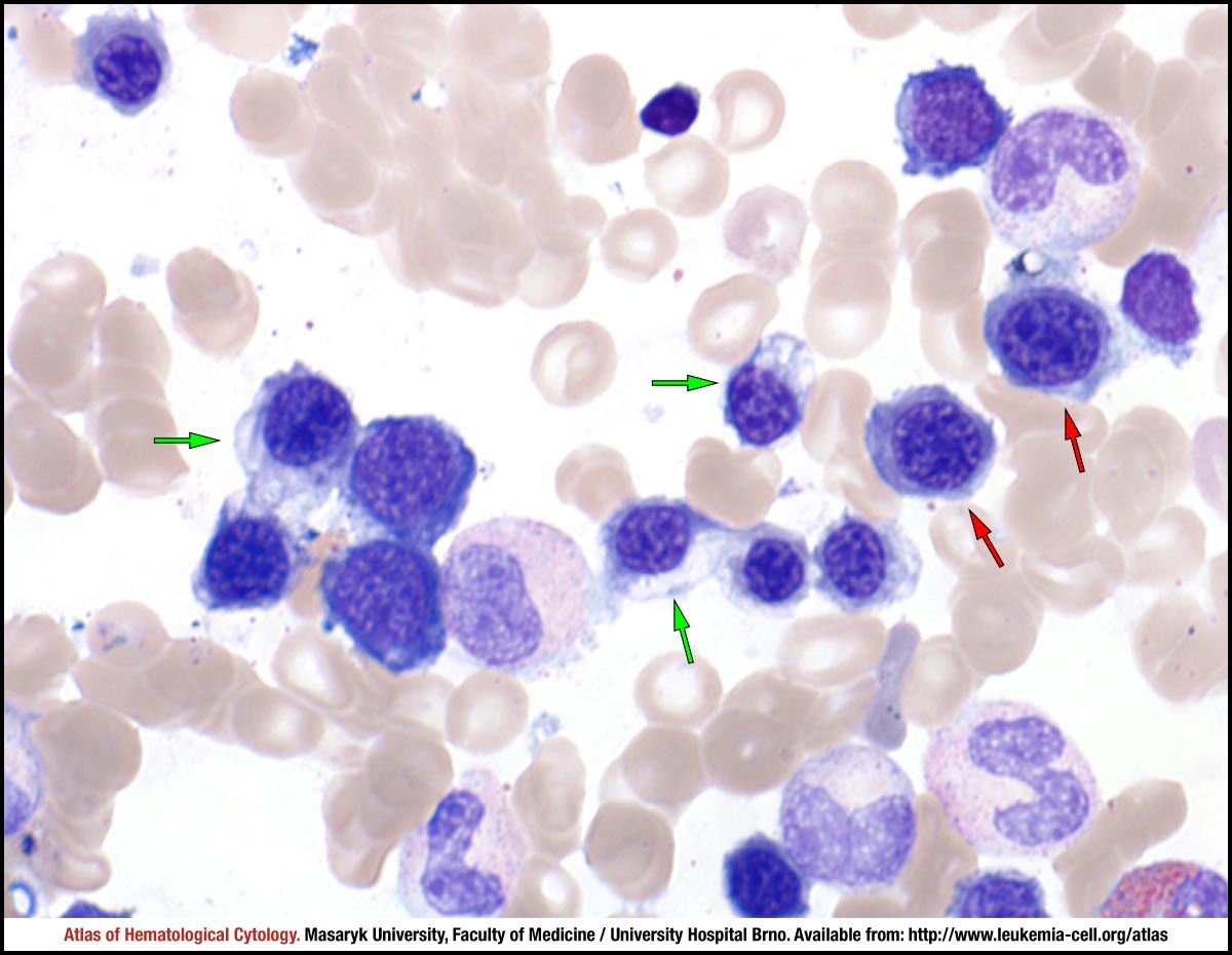 zoom picture
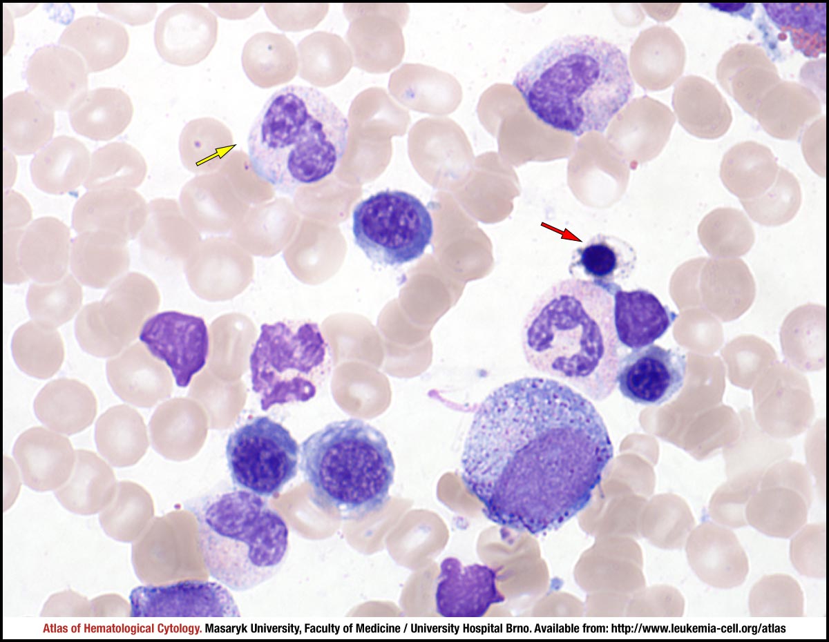 zoom picture
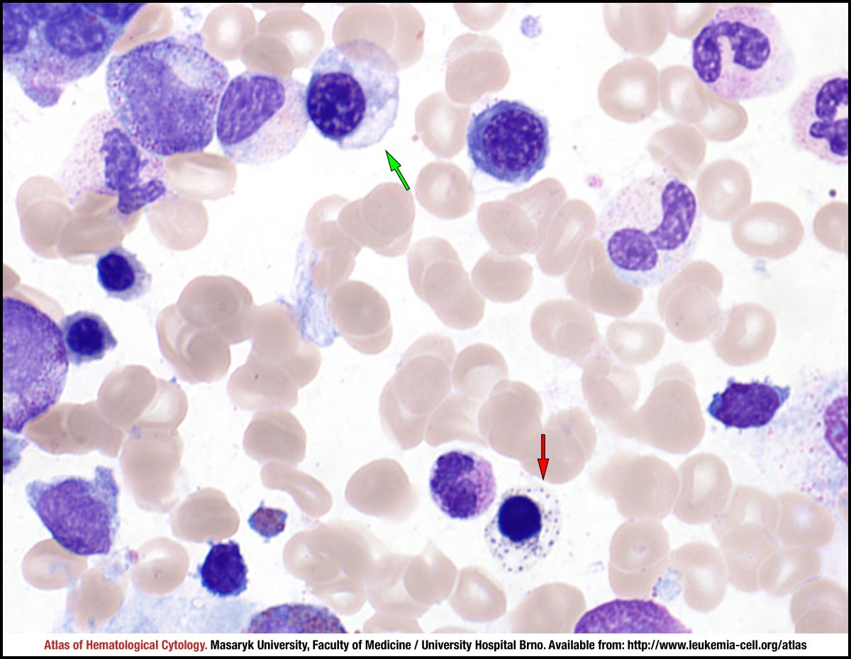 zoom picture
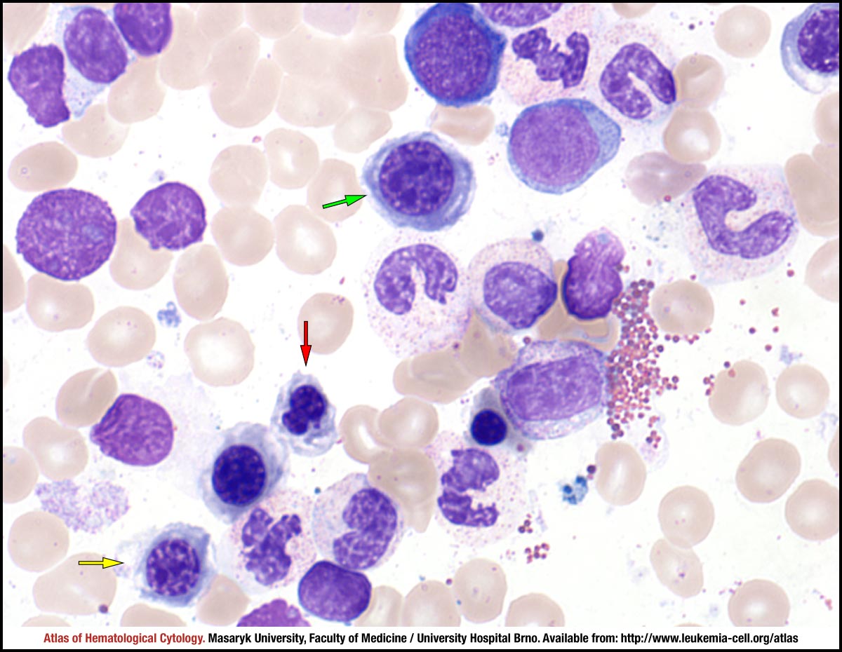 zoom picture
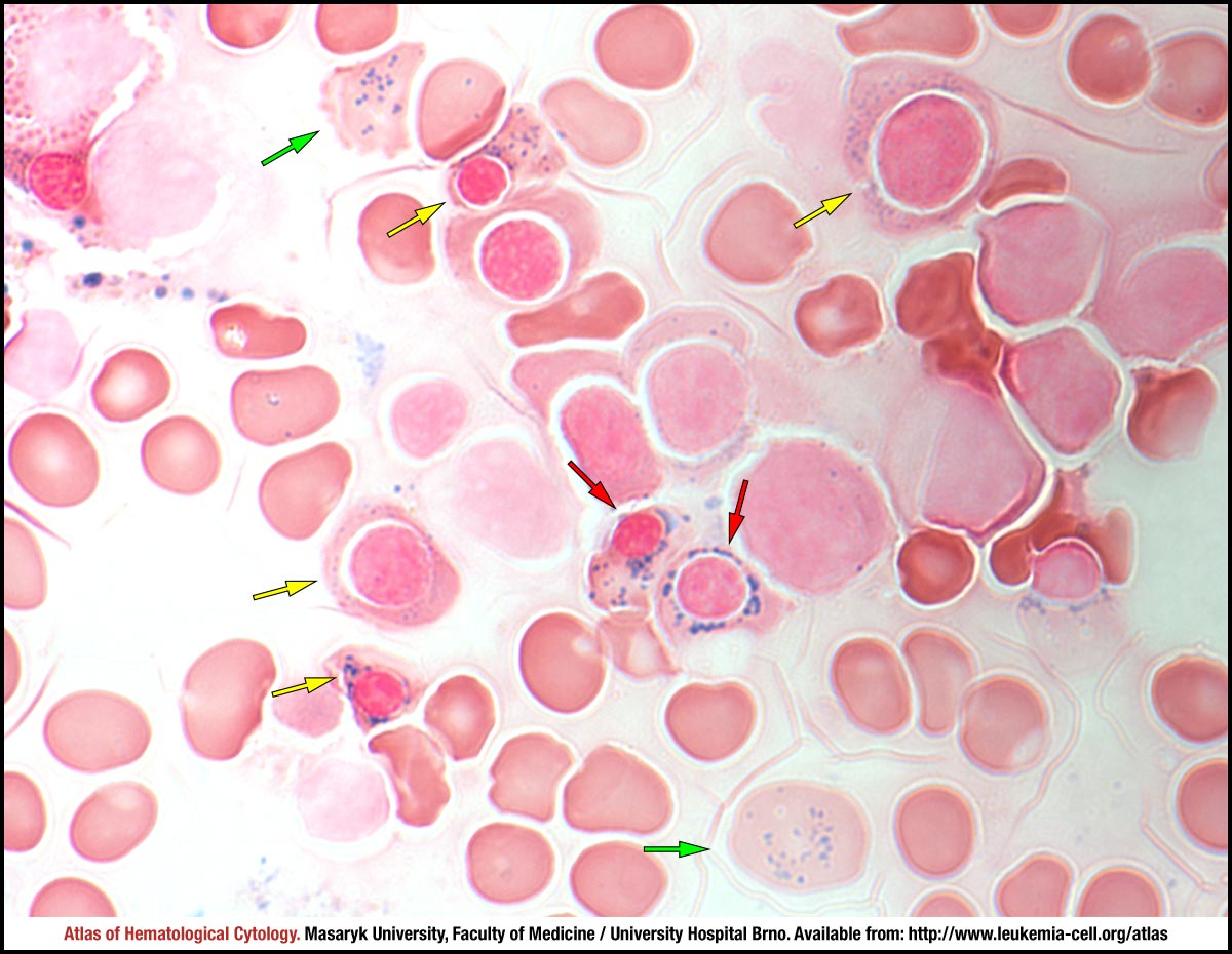 zoom picture
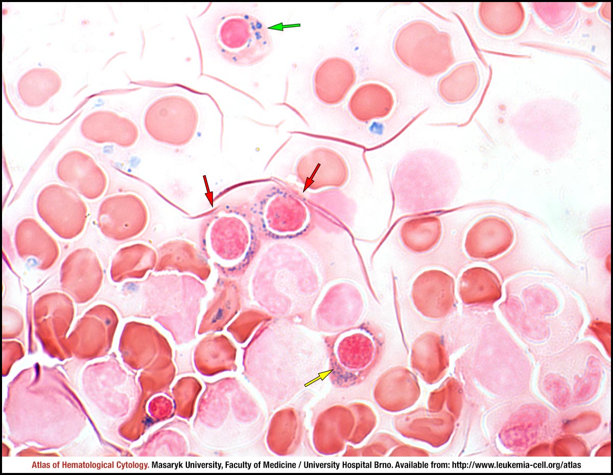 zoom picture