with flowcytometry, cytogenetic and molecular biology findings
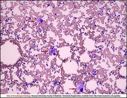
Marrow aspirate smear with an adequate cellularity and a proportional representation of haematopoietic cells. Red arrows indicate an elevated number of pathological megakaryocytes, which are usually normal to slightly decreased in size, with conspicuously non-lobulated and hypolobulated nuclei.
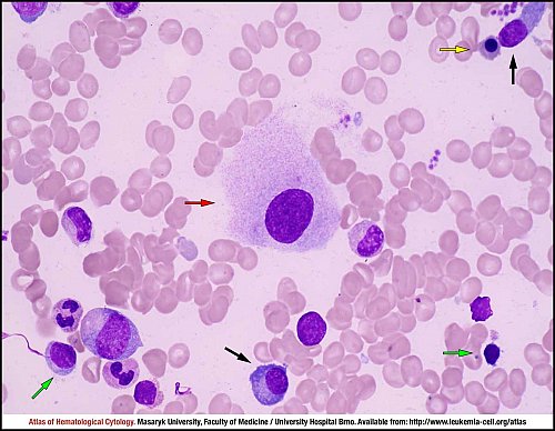
Marrow aspirate smear: one megakaryocyte with a non-lobulated nucleus (red arrow), two plasma cells (black arrows), two lymphocytes (green arrows), numerous granulocytes (unmarked) and one erythroblast (yellow arrow).
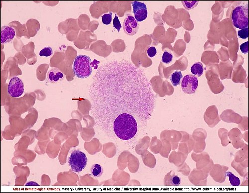
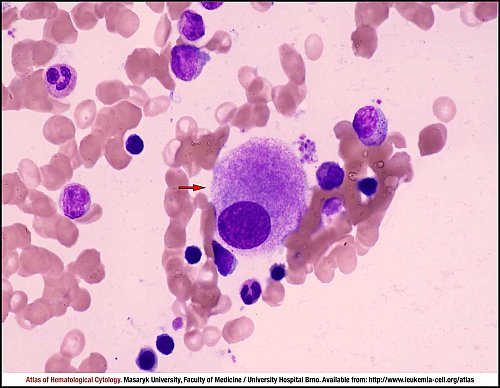
Marrow aspirate smear: one megakaryocyte with a non-lobulated nucleus (red arrow) and normal haematopoietic cells.
Marrow aspirate smear: one megakaryocyte with a non-lobulated nucleus (red arrow), normal haematopoietic cells.
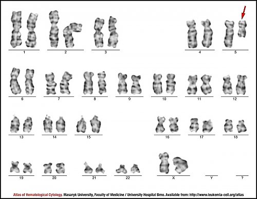
G-banded karyotype of a bone marrow metaphase cell showing 46,XX,del(5)(q13q33). The deleted chromosome 5 is indicated by a red arrow.
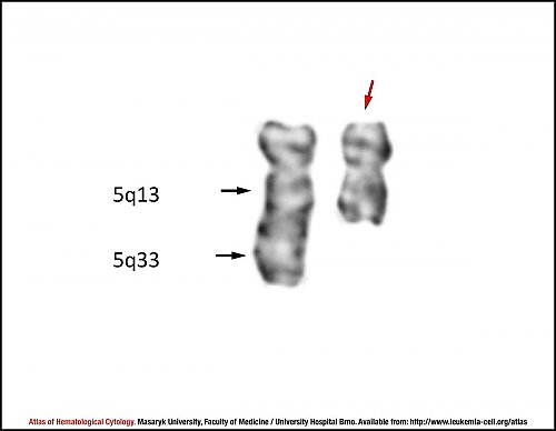
G-banded partial karyotype demonstrating an interstitial deletion in regions 5q13->5q33 (breakpoint sites are indicated by black arrows). The deleted chromosome 5 is indicated by a red arrow.
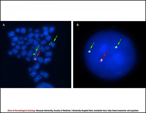
Fluorescence in situ hybridisation (FISH) of the deletion 5q31.2 performed on (A) metaphase chromosomes and (B) an interphase cell. The orange-labelled probe indicates the chromosomal region 5q31.2 (red arrows), whereas the green-labelled probe covers the region 5p15.2-p15.3 (green arrows). Only one orange signal indicates the deletion 5q31.2 in metaphase chromosomes and in the interphase cell.
Atlas of Haematological Cytology [online]. 2016 [cit. 2026-7-05]. Available from WWW: http://www.leukemia-cell.org/atlas.
2026 CELL - Atlas of Haematological Cytology | site map
 zoom picture
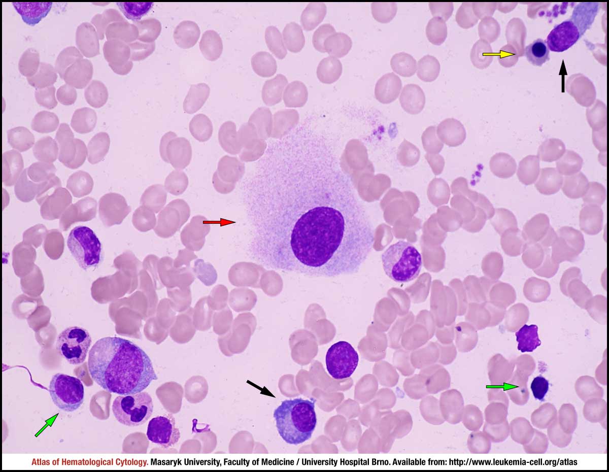 zoom picture
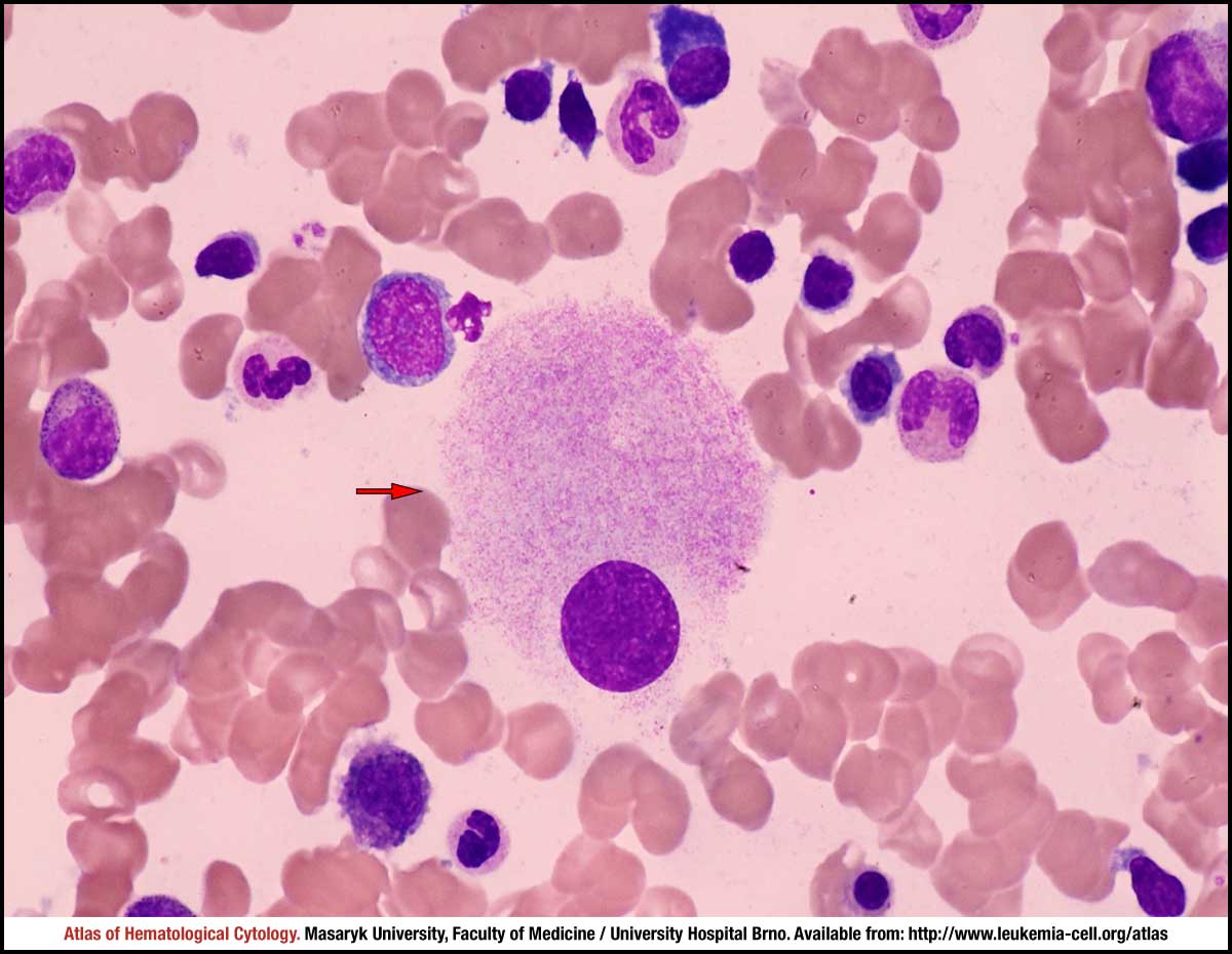 zoom picture
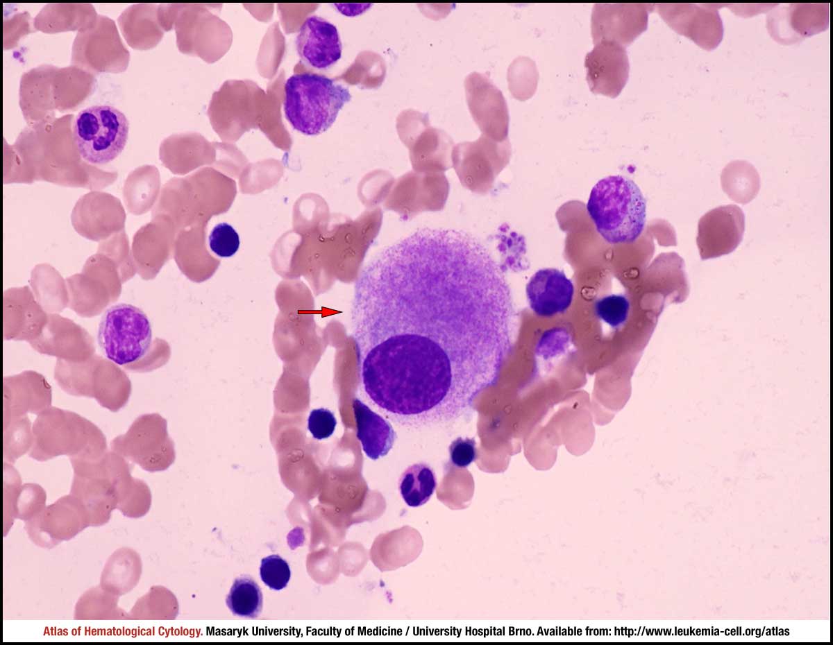 zoom picture
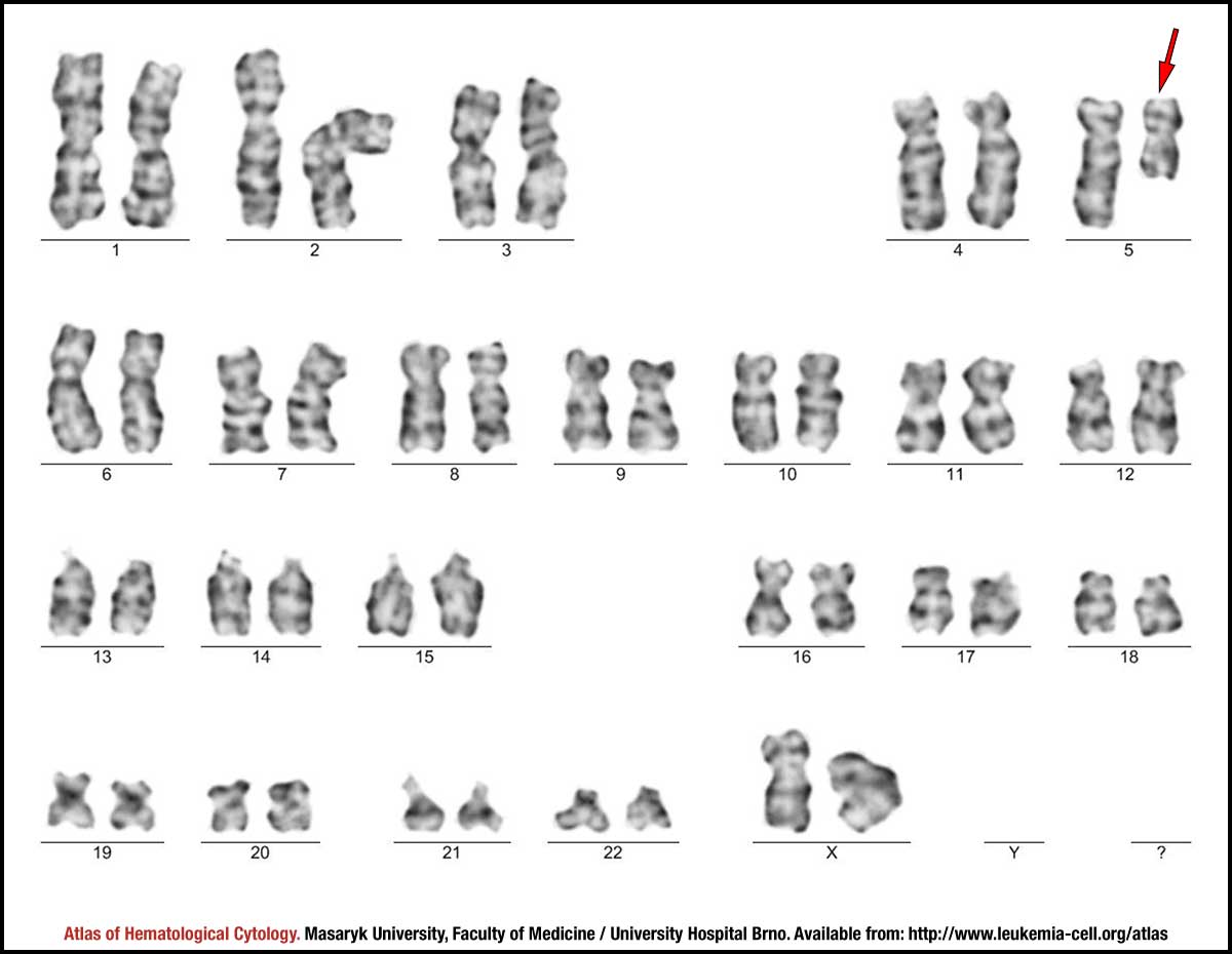 zoom picture
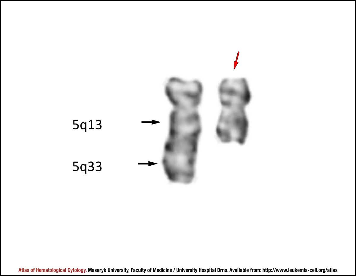 zoom picture
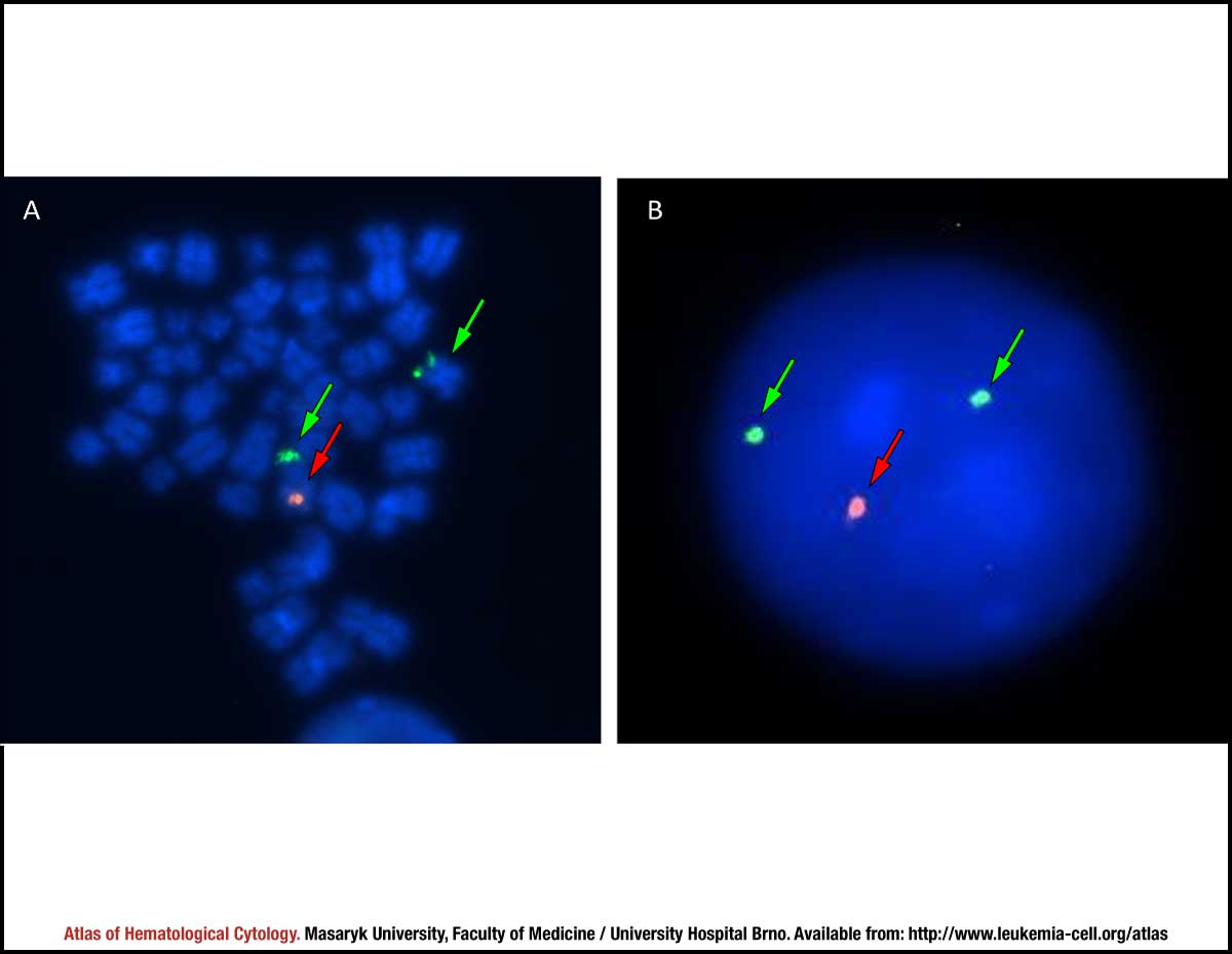 zoom picture