with flowcytometry, cytogenetic and molecular biology findings
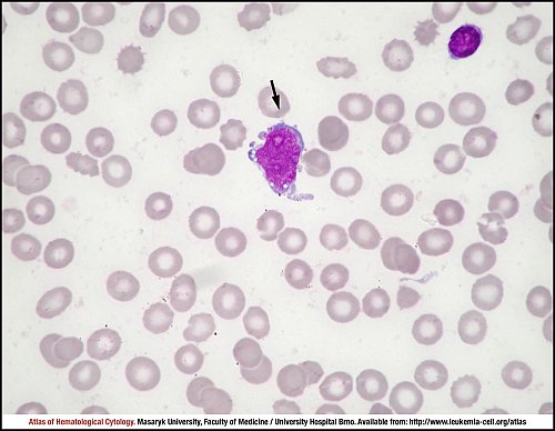
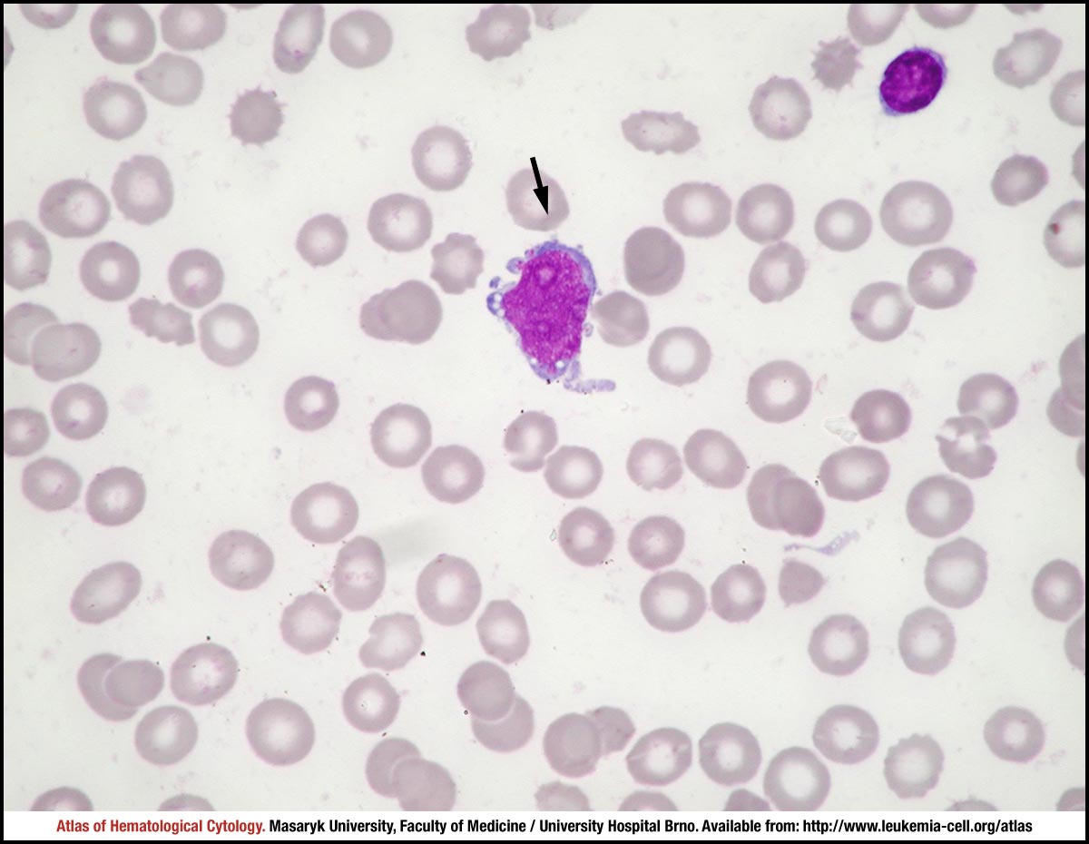
The black arrow shows a lymphoblast with a high nuclear-cytoplasmic ratio; the nucleus contains fine chromatin, three nucleoli of different sizes and irregularities in its contour, here in the form of short protrusions. The cytoplasm is moderately basophilic with fine vacuolation and fine protrusions. A mature lymphocyte is seen on the top right.
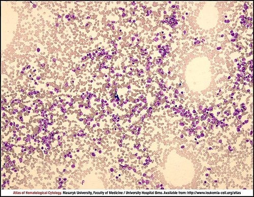
The marrow aspirate smear is rich in cells, with mononuclear lymphoblasts as the prevailing cell type.
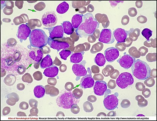
Green arrows mark neutrophilic band cells (on the left) and a myelocyte (on the centre bottom). Other cells shown in the image are lymphoblasts 16–20 µm in size, with predominantly round/oval nuclei, finely condensed chromatin, multiple nucleoli, the cytoplasm either forms a narrow edge or is distinct, homogeneously basophilic and without granules, in some places forming small budlike protrusions.
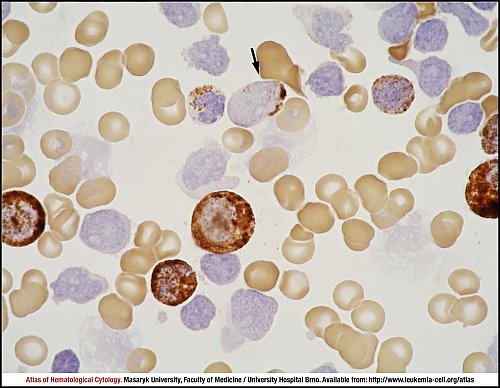
A myeloblast (containing myeloperoxidase positive granules) is marked by a black arrow; other positively staining cells correspond to neutrophils, some of them having a distinct deficit in reaction (a mature neutrophil next to the positive myeloblast). Other cells, negative for myeloperoxidase, are lymphoblasts.
see also myeloblastic crisis
Atlas of Haematological Cytology [online]. 2016 [cit. 2024-4-20]. Available from WWW: http://www.leukemia-cell.org/atlas.
2024 CELL - Atlas of Haematological Cytology | site map
 zoom picture
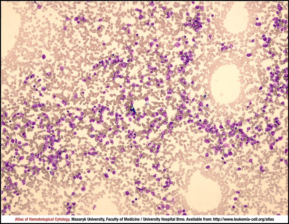 zoom picture
 zoom picture
 zoom picture