with flowcytometry, cytogenetic and molecular biology findings
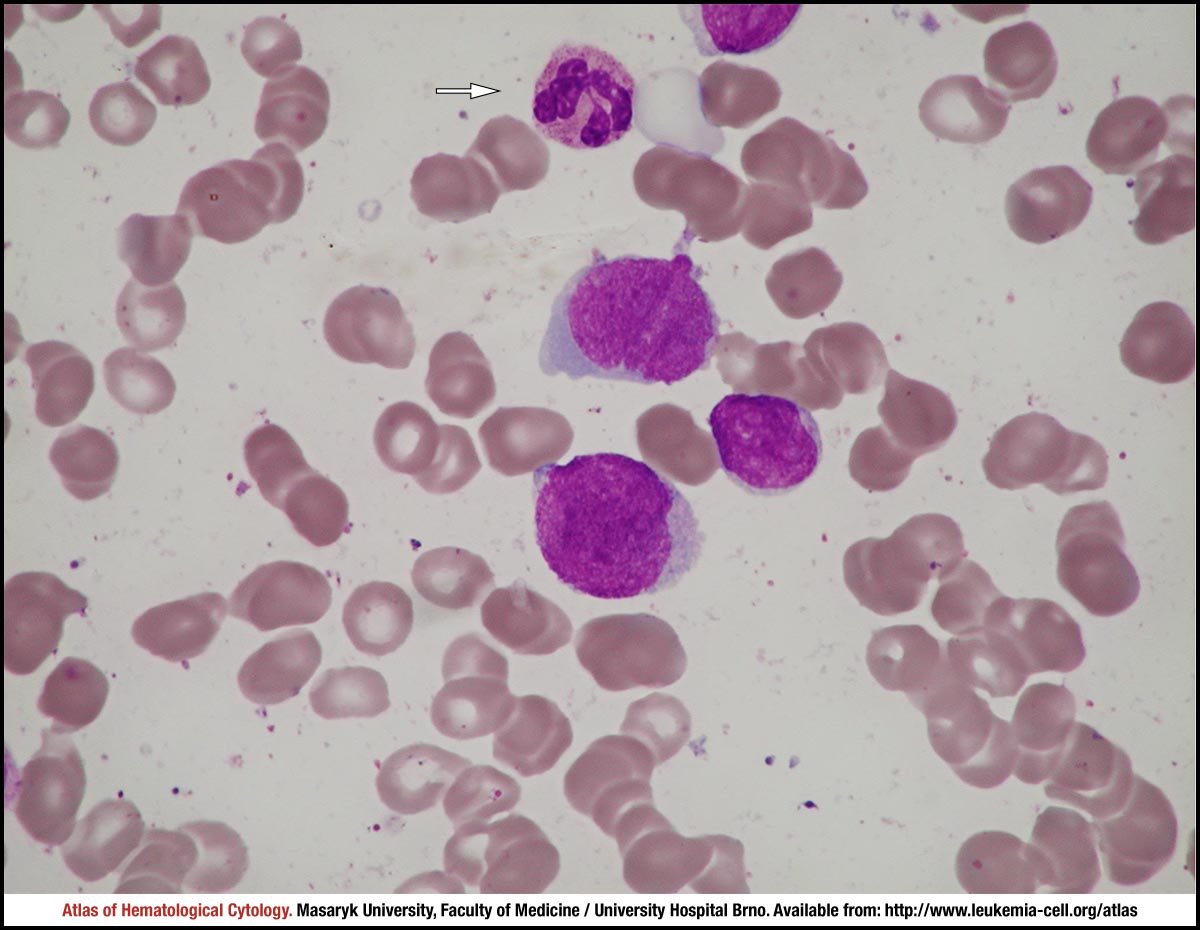
A segmented neutrophil is marked by a white arrow. The other three cells shown in the image are quite heterogeneous myeloblasts, which are different in size, with different values of nuclear-cytoplasmic ratio, with nuclei slightly indented and showing slight irregularities, containing fine to more condensed chromatin, less distinct nucleoli and sporadic irregularities in their contours (in the form of short protrusions); the cytoplasm is slightly basophilic, without granules.
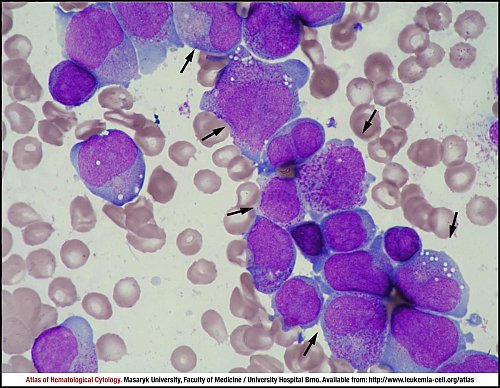
The marrow aspirate smear is rich in cells, with mononuclear cells (myeloblasts and immature granulocytes) as the prevailing cell type.
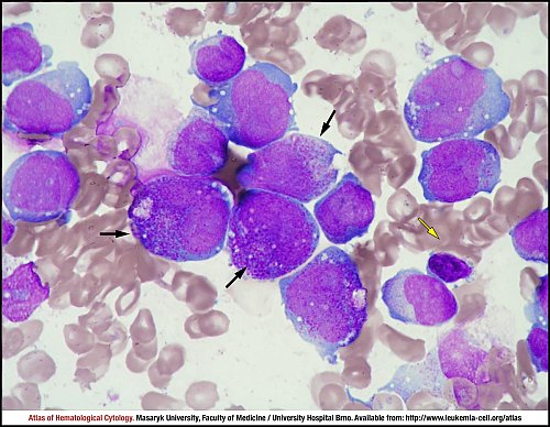
This slide is largely filled by a heterogeneous group of myeloblasts (unmarked cells), which are 16–22 µm in size. The nuclear size and shape are irregular and greatly variable, nuclei are often kidney-shaped or bilobed, contain very fine chromatin, often multiple nucleoli, their cytoplasm usually forms a broader edge, is more intensely homogeneously basophilic, without granules, with multiple azurophilic granules or with numerous vacuoles. Furthermore, large atypical promyelocytes (black arrows) and one lymphocyte (yellow arrow) are also shown in the image.
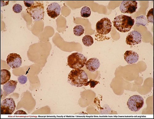
The myeloperoxidase-positive cells correspond to myeloblasts and neutrophils. The cell negative for MPO staining (black arrow) is a lymphocyte.
Atlas of Haematological Cytology [online]. 2016 [cit. 2024-4-18]. Available from WWW: http://www.leukemia-cell.org/atlas.
2024 CELL - Atlas of Haematological Cytology | site map
 zoom picture
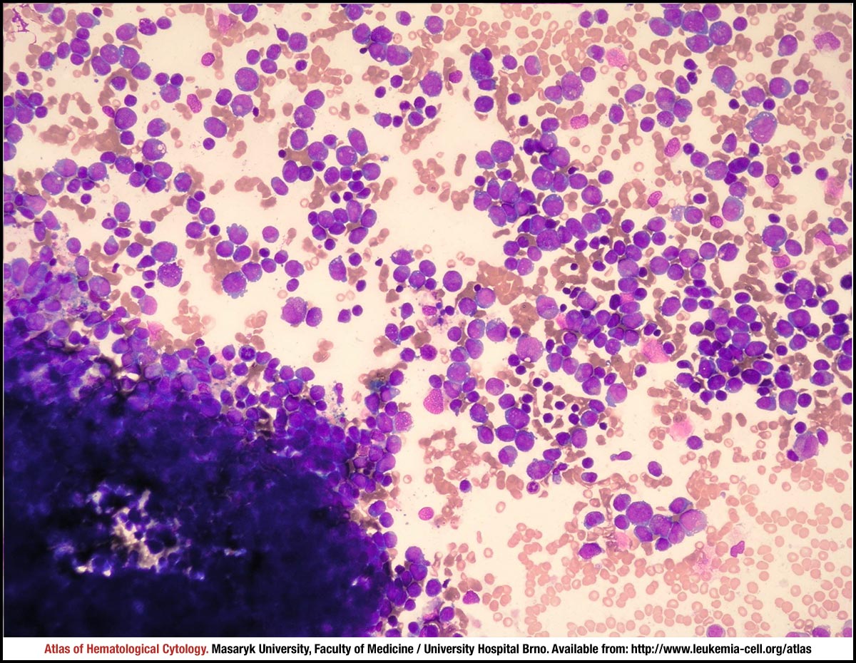 zoom picture
 zoom picture
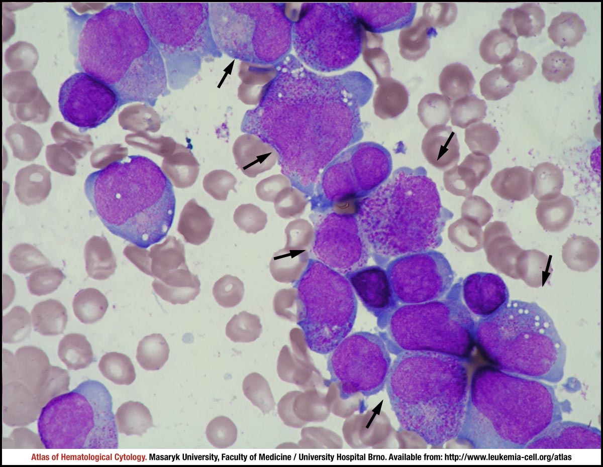 zoom picture
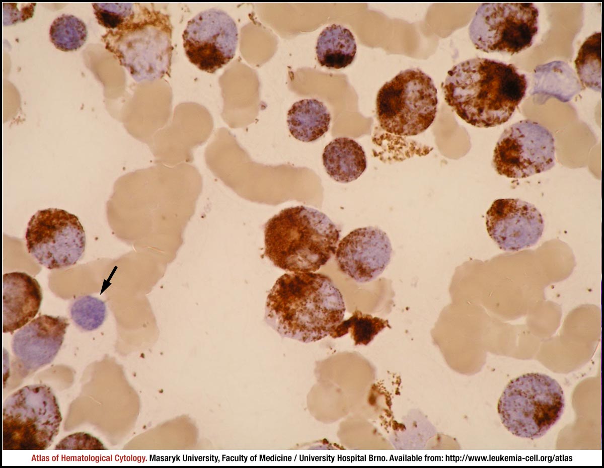 zoom picture