with flowcytometry, cytogenetic and molecular biology findings
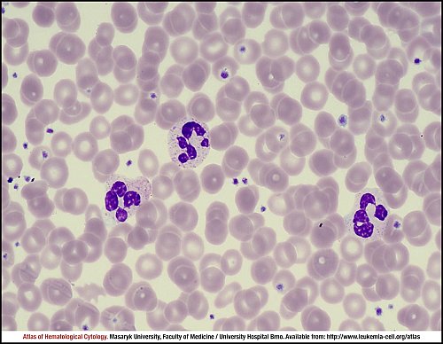
Peripheral blood smear showing a packed film appearance, three segmented neutrophils and many thrombocytes.
The marrow aspirate smear is normocellular with one bigger fragment of haematopoietic cells and with six mature megakaryocytes.
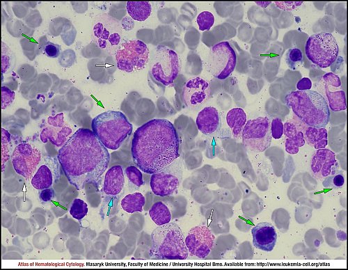
Marrow aspirate smear, granulocytic lineage with normal cellular maturation and a mild hypogranularity, three eosinophils (white arrows), a few erythroid precursors (green arrows) and two myeloblasts (blue arrows).
Marrow aspirate smear with two megakaryocytes. The giant element on the right side (white arrow) has deeply lobulated nuclei, whereas the second cell is smaller with fragments of nuclei. The picture also shows an activated plasma cell (yellow arrow).
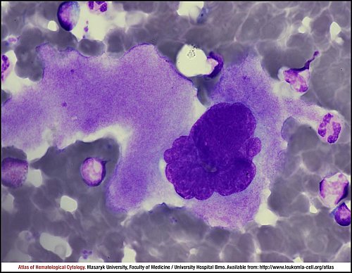
Marrow aspirate smear with a typical megakaryocyte – the giant cell with bulbous nuclei and spacious granulocytic cytoplasm.
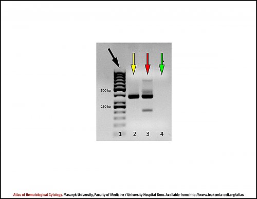
This figure shows PCR amplification products. The first band shows a JAK2-V617F-negative sample (lane 2, yellow arrow), whereas the second sample is JAK2-V617F-positive (lane 3, red arrow). The green arrow indicates no template control (lane 4, NTC). A DNA size marker was applied (lane 1, black arrow).
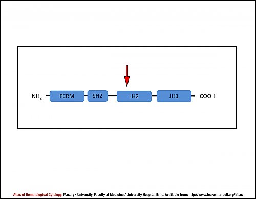
This scheme shows domains of the Jak2 protein. The mutation V617F affects the JH2 domain (red arrow), which is then unable to inhibit the JH1 domain and its tyrosin kinase signal, making it constitutively active even in the absence of ligand.
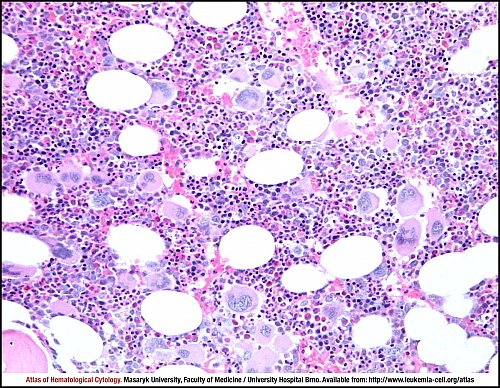
Primary polycythaemia. 60-year old man, haemoglobin 189 g/L, haematocrit 0.596, JAK2 mutated. Hypercellular bone marrow with a trilinear myeloproliferation. Increased number of megakaryocytes with a tendency to form loose clusters, enlarged cells without significant “dysplastic” features. Large clusters of left-shifted erythroid cells, maturing granulocytic series.
Bone marrow trephine biopsy, haematoxylin and eosin stain.
Atlas of Haematological Cytology [online]. 2016 [cit. 2026-7-11]. Available from WWW: http://www.leukemia-cell.org/atlas.
2026 CELL - Atlas of Haematological Cytology | site map
 zoom picture
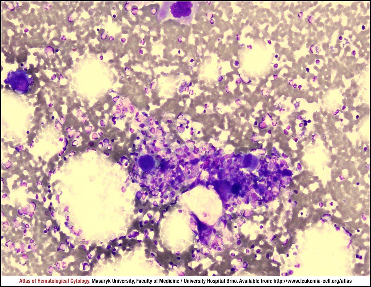 zoom picture
 zoom picture
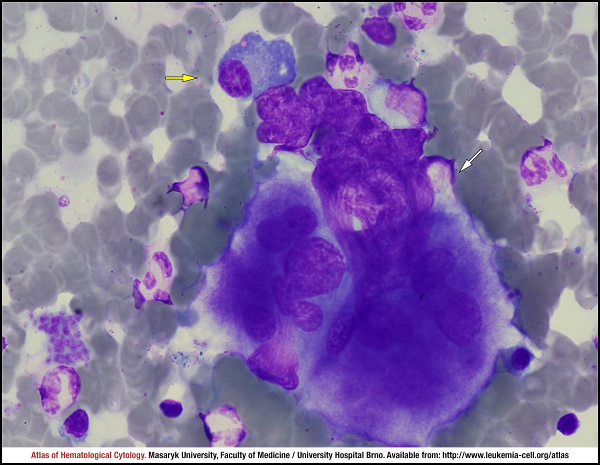 zoom picture
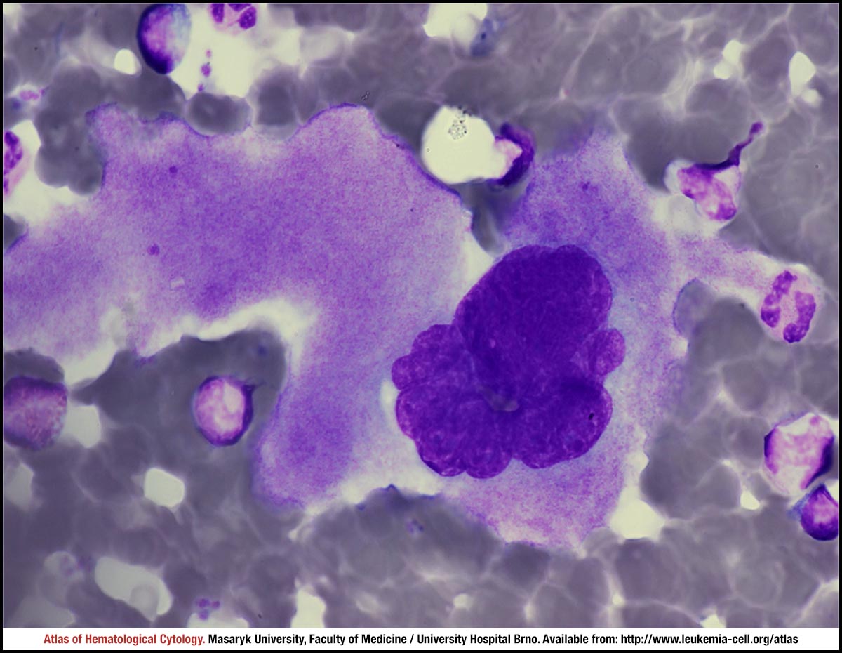 zoom picture
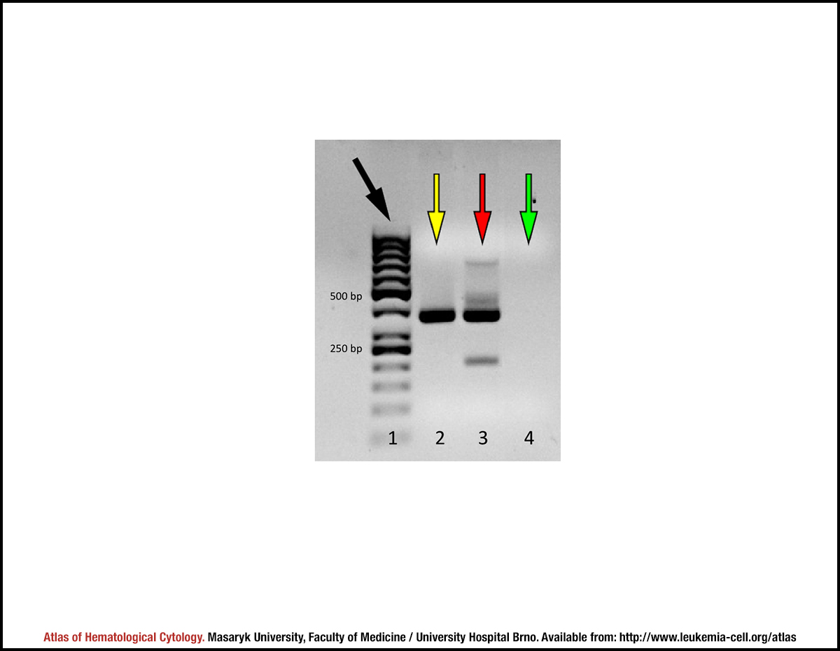 zoom picture
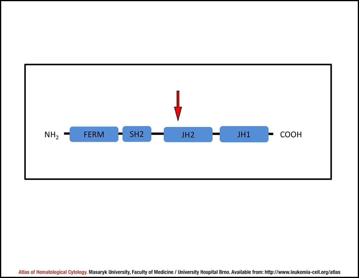 zoom picture
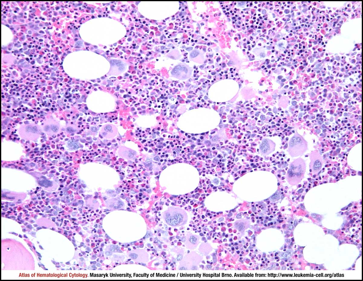 zoom picture