with flowcytometry, cytogenetic and molecular biology findings
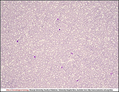
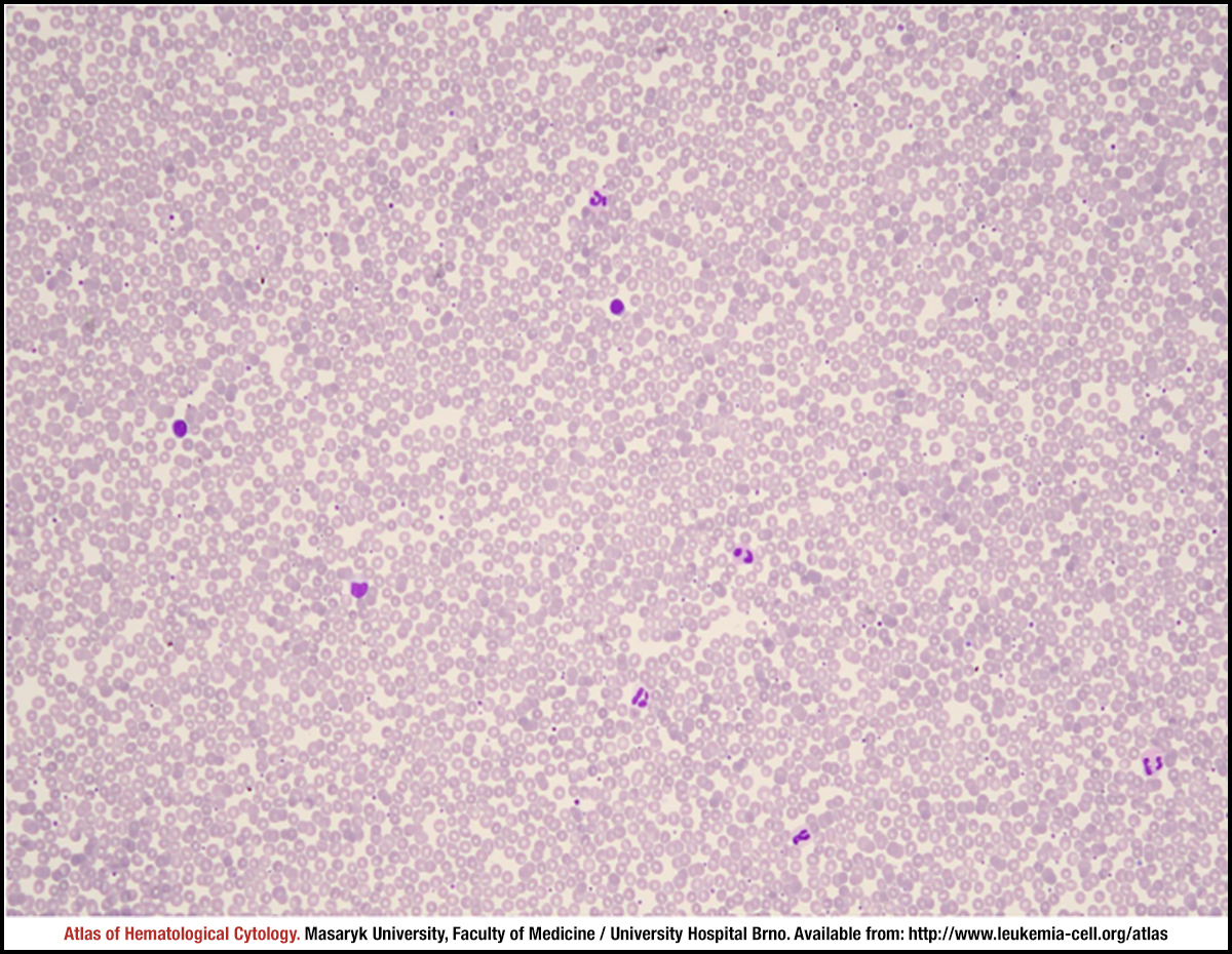
Peripheral blood smear with a normal differential white cell count.
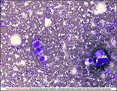
The marrow aspirate smear is hypercellular with an increasing number of megakaryocytes (six cells on the photograph).
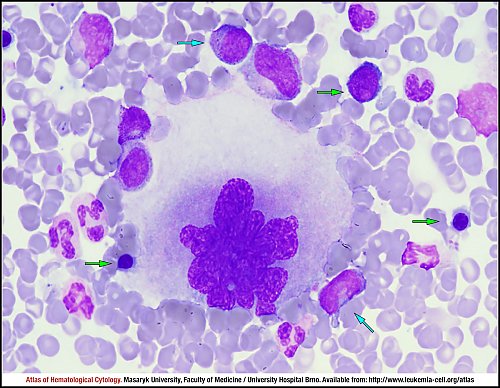
In this marrow aspirate smear, one megakaryocyte is enlarged with bulbous nuclei and abnormal chromatin patterns of chromatin clumping. There are three erythroid precursors (green arrows), a few granulocytes and two granular and agranular myeloblasts (blue arrows).
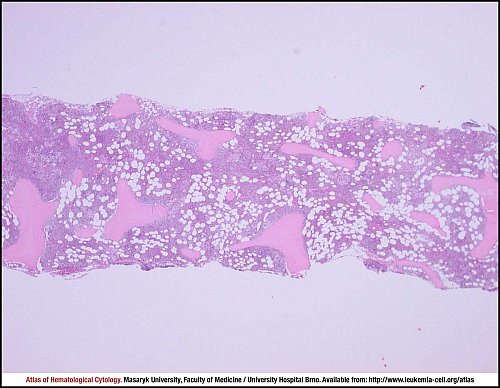
Primary myelofibrosis, early fibrosis. 65-year old woman, leucocytosis 12×109/L, thrombocytosis 1,000×109/L, JAK2 mutated. Hypercellular haematopoiesis.
Bone marrow trephine biopsy, haematoxylin and eosin stain.
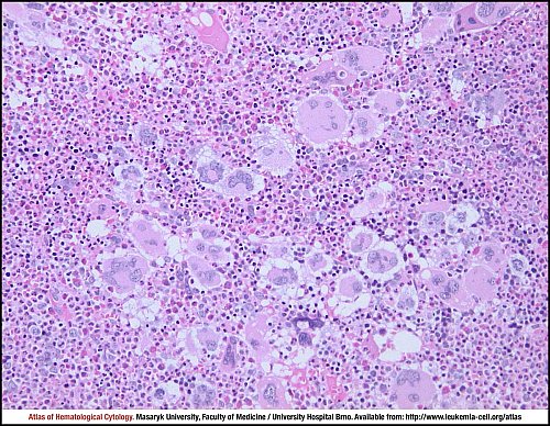
Primary myelofibrosis, early fibrosis. 65-year old woman, leucocytosis 12×109/L, thrombocytosis 1,000×109/L, JAK2 mutated. Trilinear myeloproliferation with a conspicuous increase of megakaryopoiesis.
Bone marrow trephine biopsy, haematoxylin and eosin stain.
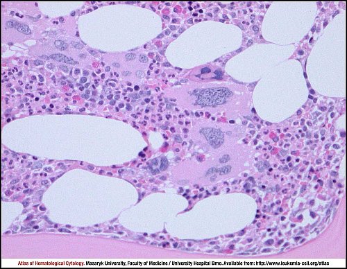
Primary myelofibrosis, early fibrosis. 65-year old woman, leucocytosis 12×109/L, thrombocytosis 1,000×109/L, JAK2 mutated. Megakaryocytes are forming dense, “cohesive” clusters. Enlarged cells often have hypolobated, “cloud-like” nuclei and variable staining of cytoplasm.
Bone marrow trephine biopsy, haematoxylin and eosin stain.
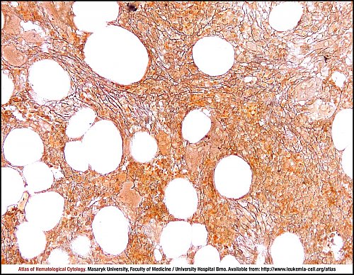
Primary myelofibrosis, early fibrosis. 65-year old woman, leucocytosis 12×109/L, thrombocytosis 1,000×109/L, JAK2 mutated. Fine reticulin fibrosis (MF-1 according to the EUMNET classification).
Bone marrow trephine biopsy, Gomori silver impregnation stain.
Atlas of Haematological Cytology [online]. 2016 [cit. 2026-6-25]. Available from WWW: http://www.leukemia-cell.org/atlas.
2026 CELL - Atlas of Haematological Cytology | site map
 zoom picture
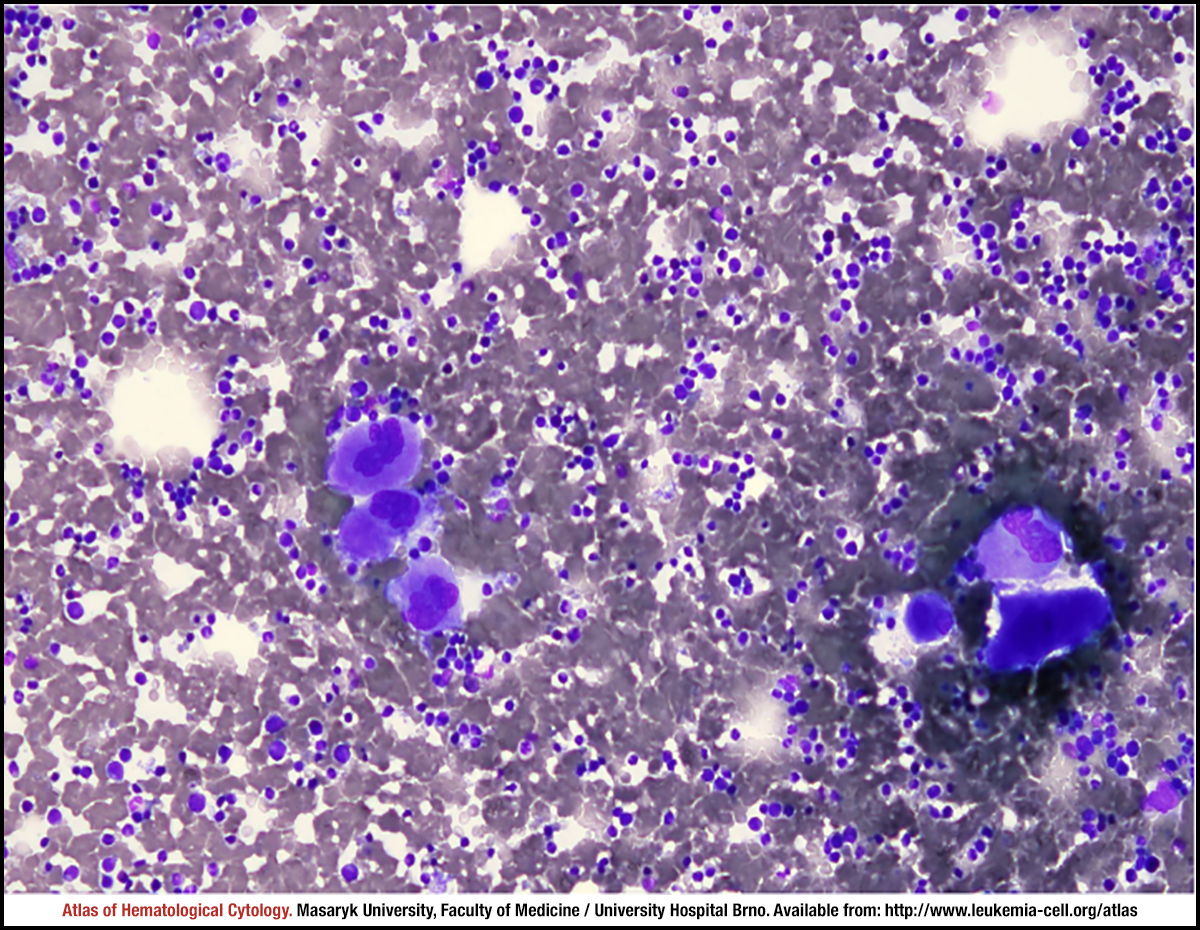 zoom picture
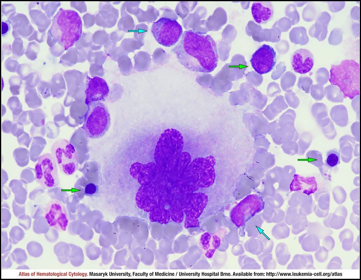 zoom picture
 zoom picture
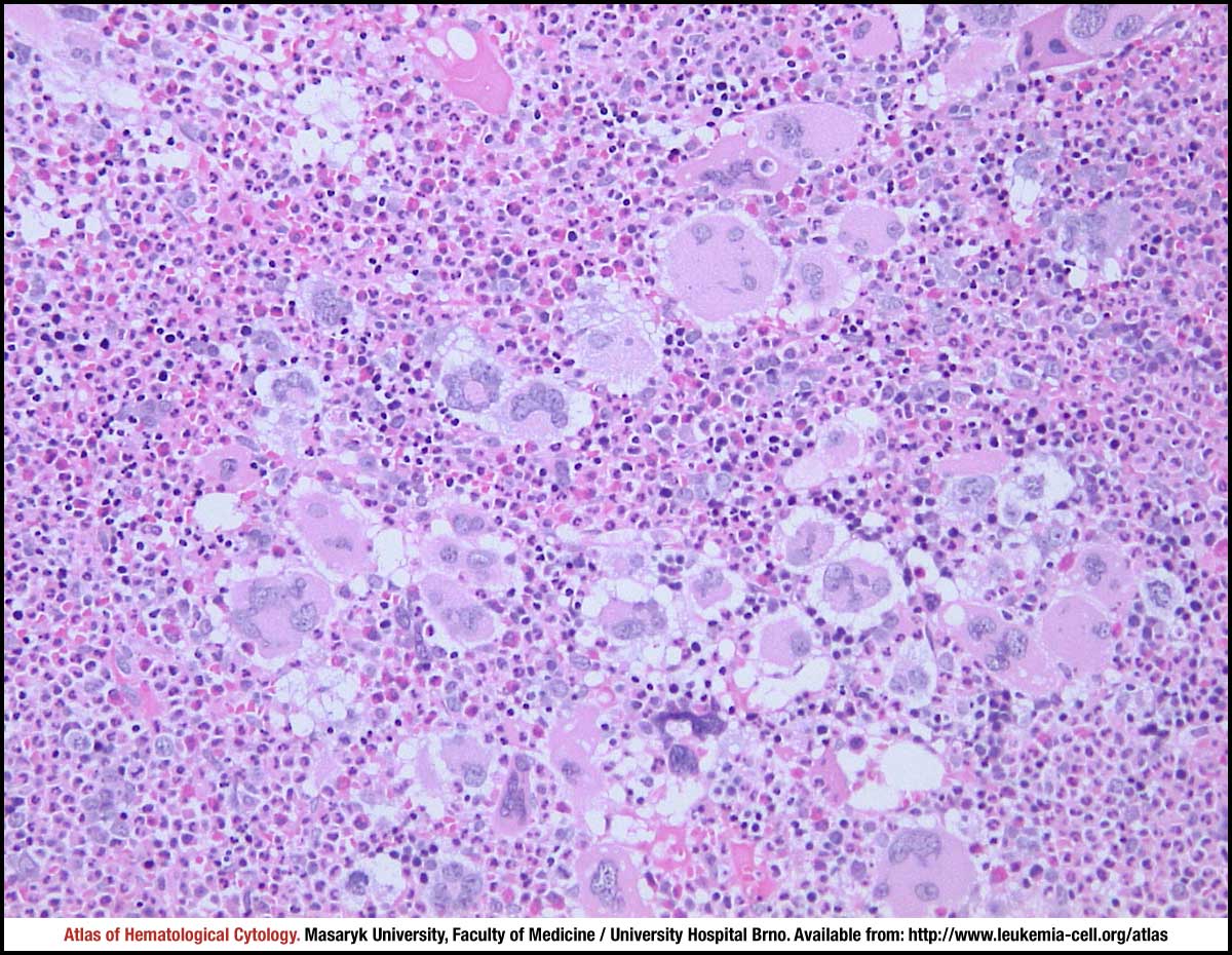 zoom picture
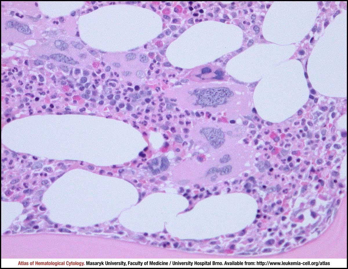 zoom picture
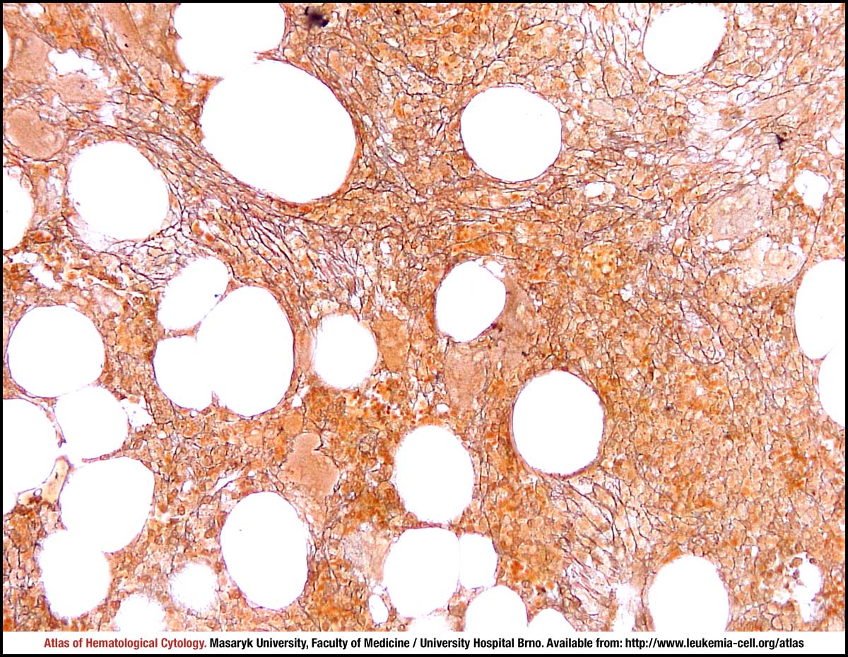 zoom picture