with flowcytometry, cytogenetic and molecular biology findings
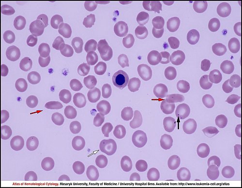
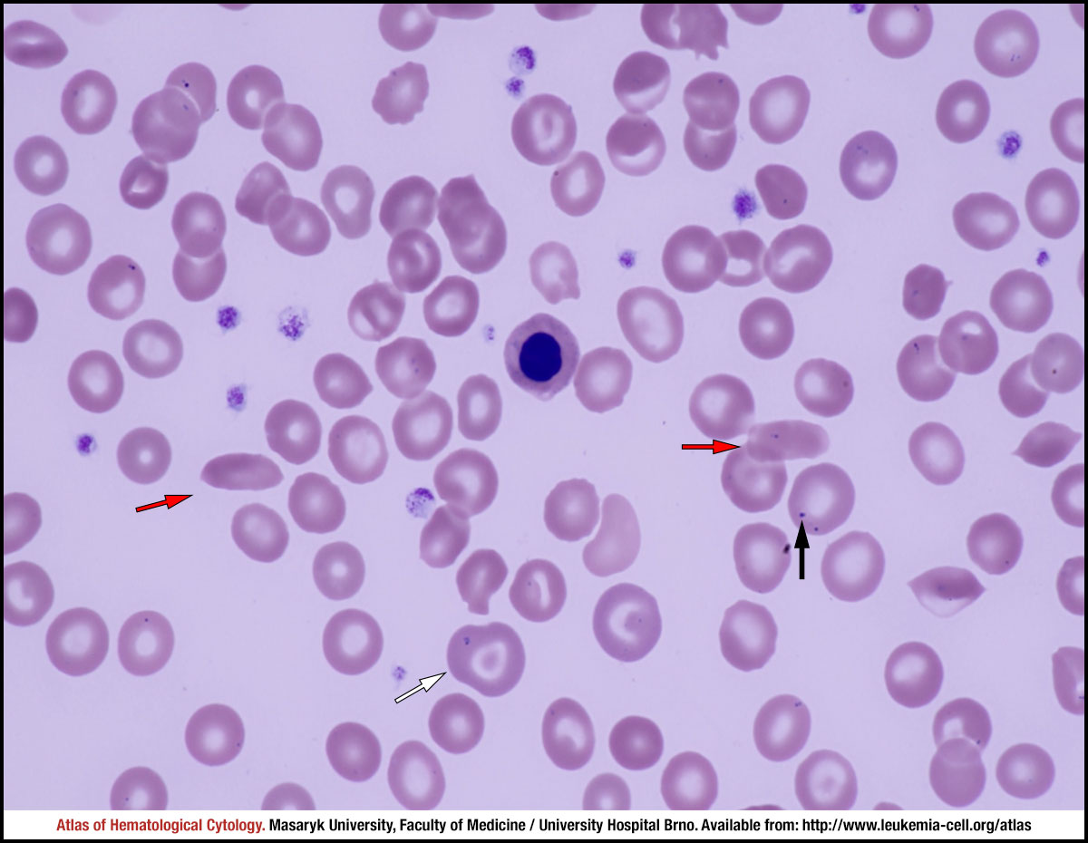
Marked anisopoikilocytosis, hypochromia and markedly atypical erythrocytes in terms of shape (numerous target cells, e.g. an erythrocyte marked by a white arrow). Sickle cells are marked by red arrows. A nucleated red blood cell – an erythroblast with a pyknotic nucleus – is shown in the middle of the image. Pappenheimer bodies, i.e. inclusion bodies composed of ferritin aggregates, are evident in some erythrocytes (marked by a black arrow).
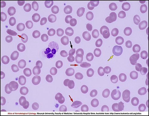
Marked anisopoikilocytosis, hypochromia and markedly atypical erythrocytes in terms of shape. Sickle cells are marked by red arrows. A segmented neutrophil is shown in the middle of the image. An erythrocyte containing a Pappenheimer body is marked by a black arrow, and an early polychromatophilic erythroblast – a reticulocyte – is marked by a yellow arrow.
Marked anisopoikilocytosis, hypochromia and markedly atypical erythrocytes in terms of shape. Sickle cells are marked by red arrows. A segmented neutrophil is shown in the middle of the image. An erythrocyte containing a Howell–Jolly body is marked by a green arrow, and a mechanically damaged erythrocyte – a keratocyte – is marked by a cyan arrow.
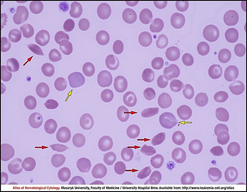
Marked anisopoikilocytosis, hypochromia and markedly atypical erythrocytes in terms of shape. Sickle cells are marked by red arrows, and reticulocytes are marked by yellow arrows.
Atlas of Haematological Cytology [online]. 2016 [cit. 2025-7-19]. Available from WWW: http://www.leukemia-cell.org/atlas.
2025 CELL - Atlas of Haematological Cytology | site map
 zoom picture
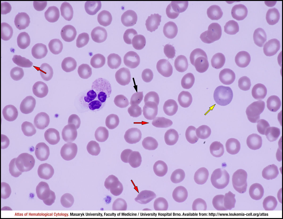 zoom picture
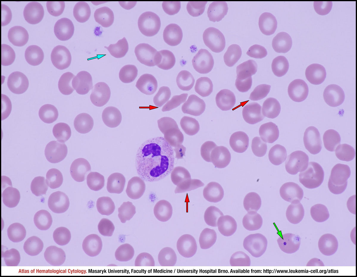 zoom picture
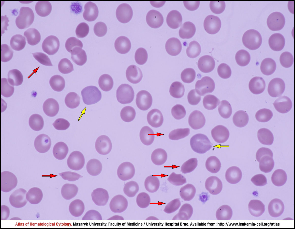 zoom picture