with flowcytometry, cytogenetic and molecular biology findings
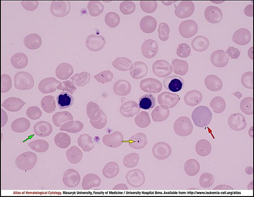
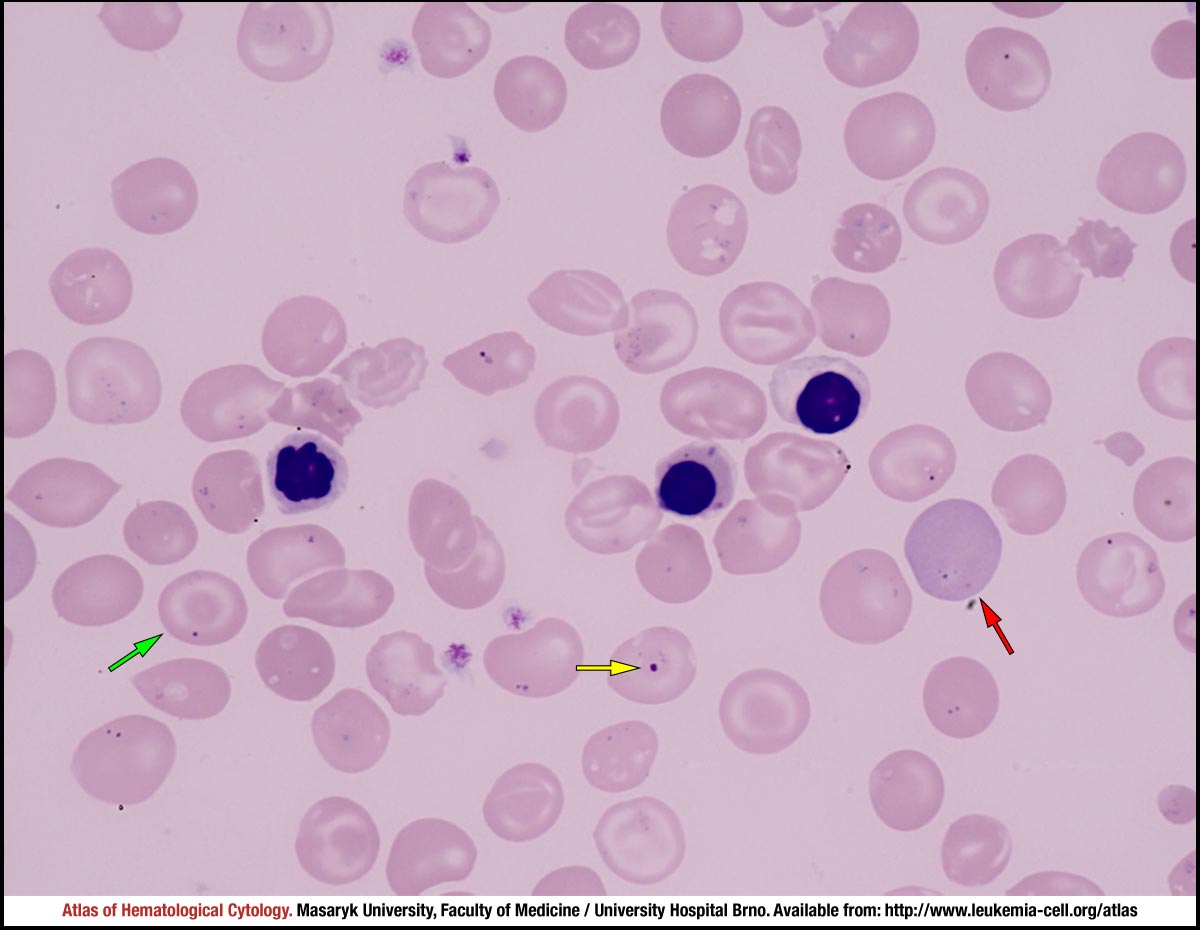
Marked anisopoikilocytosis, anisohypochromia and markedly atypical erythrocytes (in terms of shape) are shown in this peripheral blood smear: for example, target cells (green), polychromasia (red) and, in the centre of the image, three oxyphilic erythroblasts containing pyknotic nuclei with irregular contours. A Howell–Jolly body is marked by a yellow arrow.
Atlas of Haematological Cytology [online]. 2016 [cit. 2025-7-14]. Available from WWW: http://www.leukemia-cell.org/atlas.
2025 CELL - Atlas of Haematological Cytology | site map
 zoom picture