with flowcytometry, cytogenetic and molecular biology findings
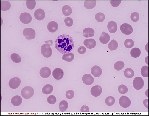
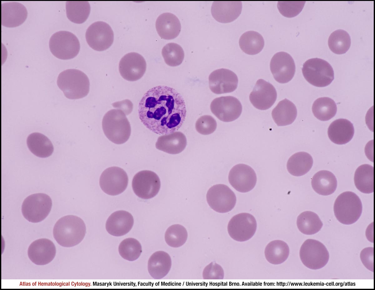
Macrocytosis of erythrocytes, numerous ovalocytes, sporadically spherocytes. A hypersegmented neutrophil is shown in the centre of the image – a finding typical for this diagnosis.
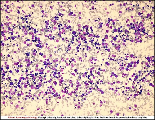
The typically “blue” marrow is indicative of the presence of less mature erythroblasts; on the whole, erythropoiesis predominates in this smear.
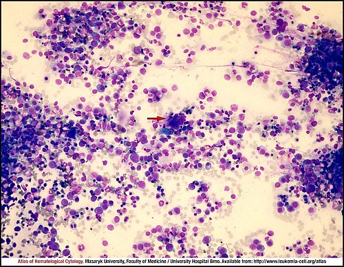
This smear shows a typically “blue” marrow, haematopoiesis is more often present in clusters, less mature erythroblasts are shown as marked megaloblasts. A large hypersegmented megakaryocyte is marked by a red arrow, and close to it (left bottom) is a macrophage with bluish cytoplasm, probably a siderophage.
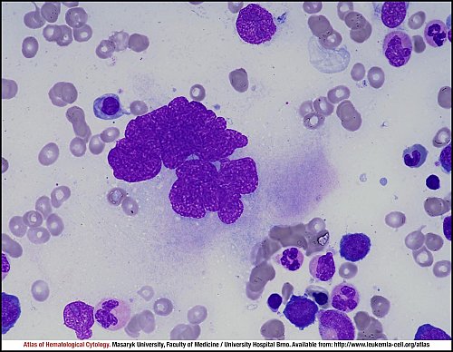
A large hypersegmented megakaryocyte is shown in the centre of the image, with typically extended chromatin.
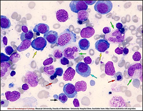
Almost the entire erythropoiesis is of megaloblastic character: the image shows proerythroblasts (yellow arrows), a basophilic erythroblast (blue arrow), a polychromatophilic erythroblast (black arrow), an orthochromatophilic erythroblast (red arrow), and megaloblastically altered neutrophilic metamyelocytes (green arrows).
Almost the entire erythropoiesis is of megaloblastic character (see also the preceding image). Megaloblastically altered erythroblasts and neutrophils are shown in particular.
Atlas of Haematological Cytology [online]. 2016 [cit. 2024-4-26]. Available from WWW: http://www.leukemia-cell.org/atlas.
2024 CELL - Atlas of Haematological Cytology | site map
 zoom picture
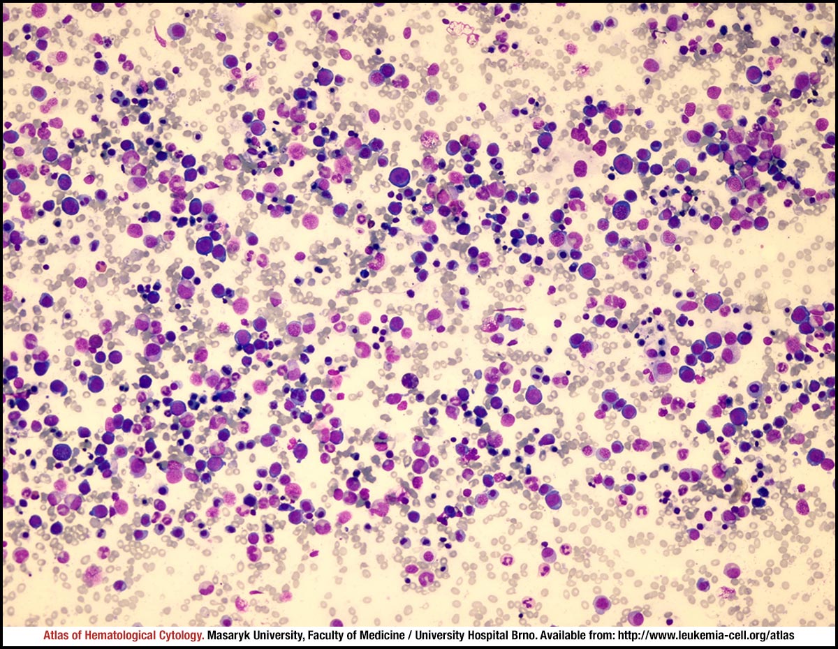 zoom picture
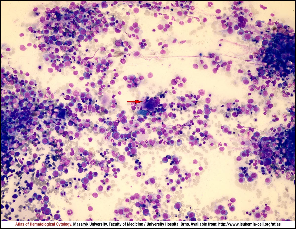 zoom picture
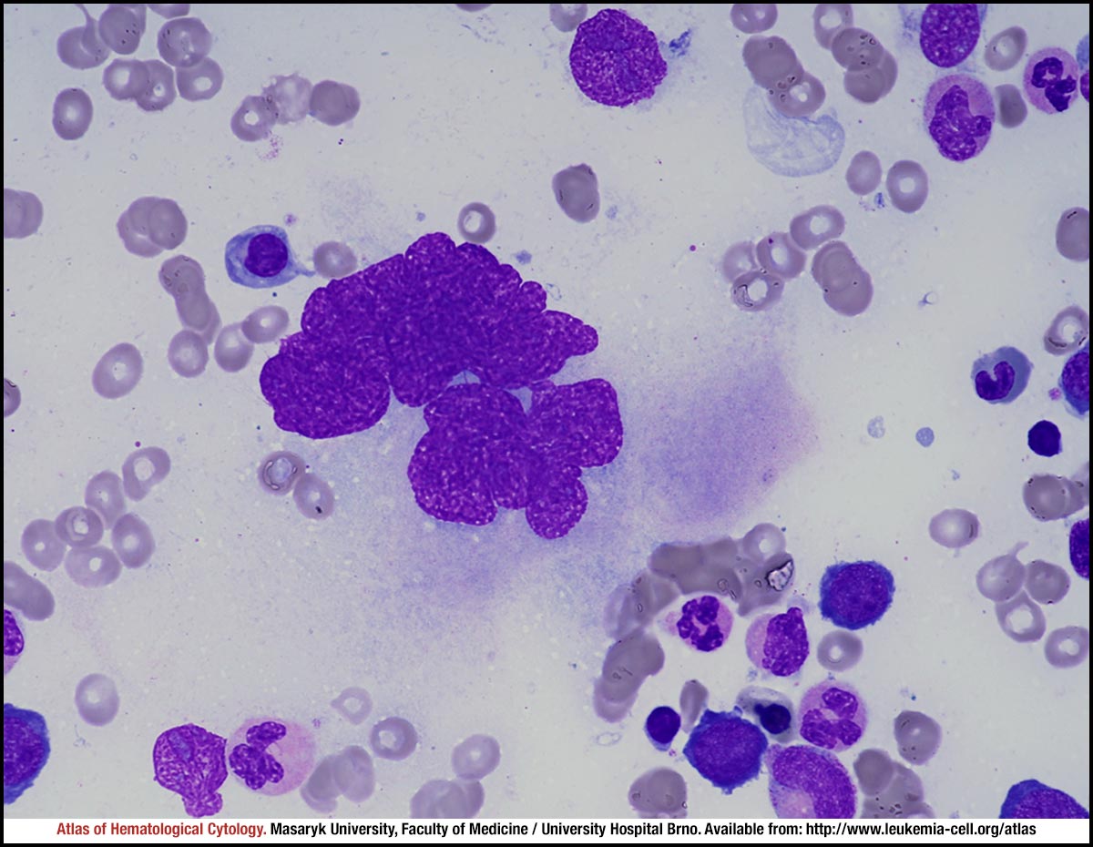 zoom picture
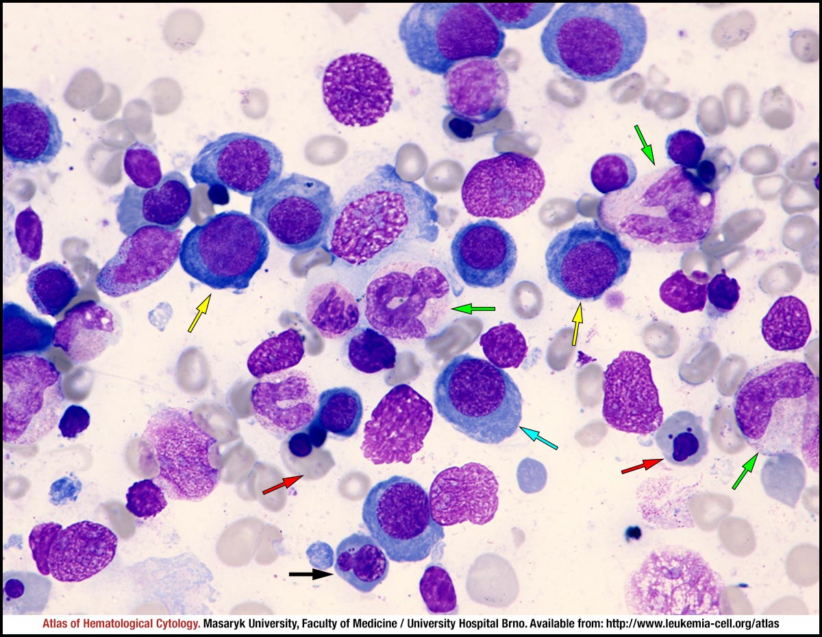 zoom picture
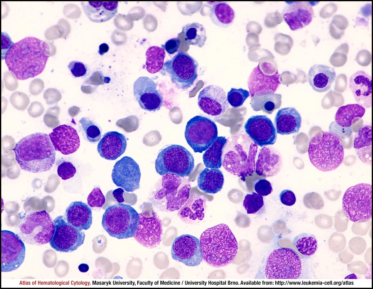 zoom picture