with flowcytometry, cytogenetic and molecular biology findings
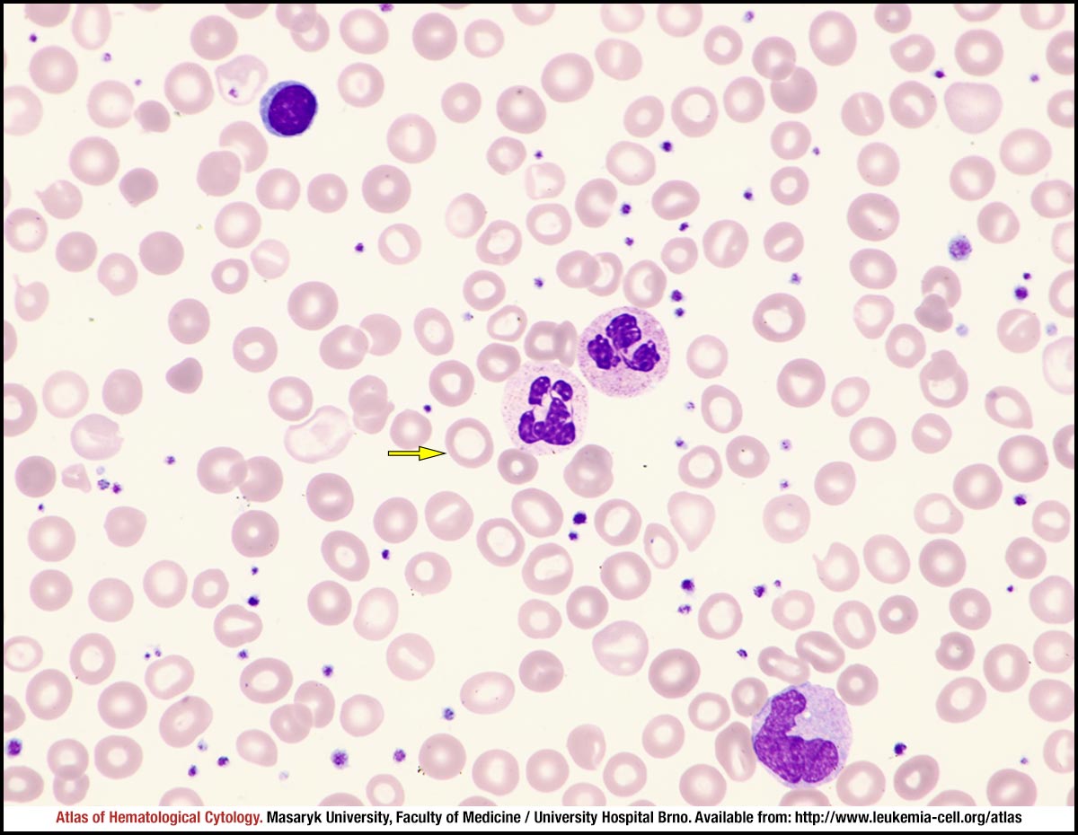
Anisomicrocytosis of erythrocytes, with many erythrocytes being so hypochromic that most of them look like anulocytes; leptocytes (yellow arrow) – erythrocytes with marked central pallor – are also occasionally present. Borderline hypersegmented neutrophils are shown in the centre of the image, a lymphocyte at the top and a monocyte at the bottom of the image.
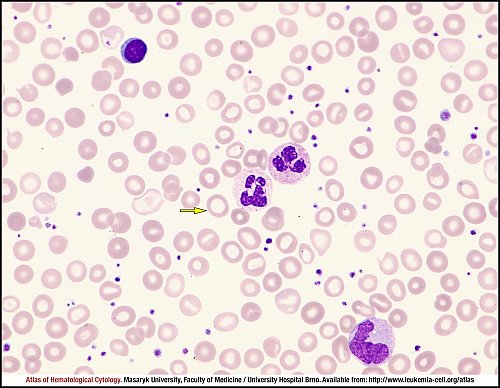
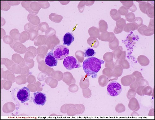
Polychromatophilic erythroblasts (yellow arrows) with impaired haemoglobinisation are showing a lack of cytoplasm. The blast element marked by a red arrow is more difficult to classify: judging by the cytoplasmic extension, it is probably a megakaryoblast. The other elements are neutrophils.
Atlas of Haematological Cytology [online]. 2016 [cit. 2024-4-25]. Available from WWW: http://www.leukemia-cell.org/atlas.
2024 CELL - Atlas of Haematological Cytology | site map
 zoom picture
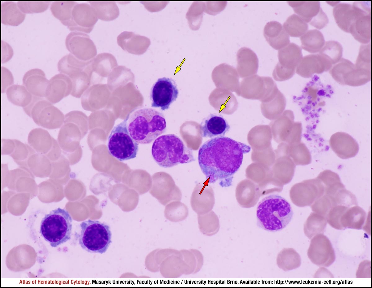 zoom picture