with flowcytometry, cytogenetic and molecular biology findings
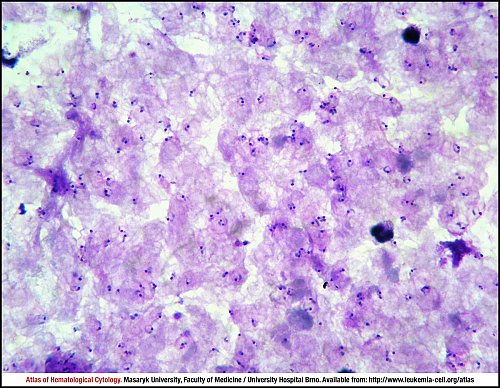
Many rings are seen in the thick blood smear. Typically only young trophozoites and gametocytes are present in the peripheral blood.
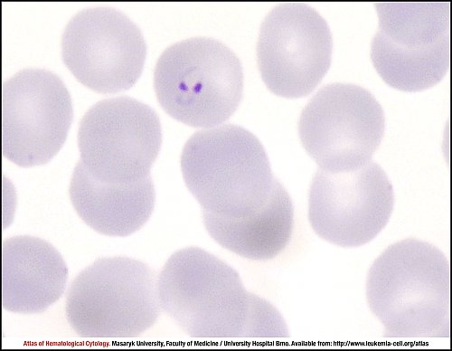
Rings in a thin blood smear show double chromatin dots, the so-called “headphones”. The infected erythrocyte is not enlarged and pigment is not present.
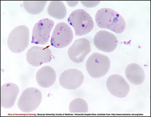
More rings in a thin blood smear. The red arrow shows an “appliqué” form, i.e. the ring is located on the periphery of the erythrocyte. Multiple infections of erythrocytes are also present. Maurer’s clefts on infected erythrocytes are visible.
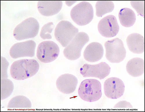
More erythrocytes infected with Plasmodium falciparum in a thin blood smear. Several “appliqué” forms are present. The red arrow shows a developing trophozoite. Maurer’s clefts are often seen in older rings.
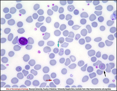
Several rings and one trophozoite are shown in this image. The red arrow shows an “appliqué” ring form, the green arrow marks two “headphone” rings and the black arrow points at a trophozoite. Infected erythrocytes are not enlarged and stippling is not seen.
Red arrows show three gametocytes in a thick blood smear. Plasmodium falciparum gametocytes are crescent- or sausage-shaped.
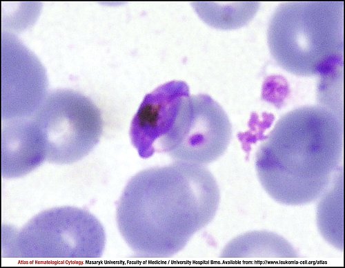
A macrogametocyte in a thin blood smear. Note the membrane of the infected erythrocyte and the dark pigment.
Atlas of Haematological Cytology [online]. 2016 [cit. 2024-5-02]. Available from WWW: http://www.leukemia-cell.org/atlas.
2024 CELL - Atlas of Haematological Cytology | site map
 zoom picture
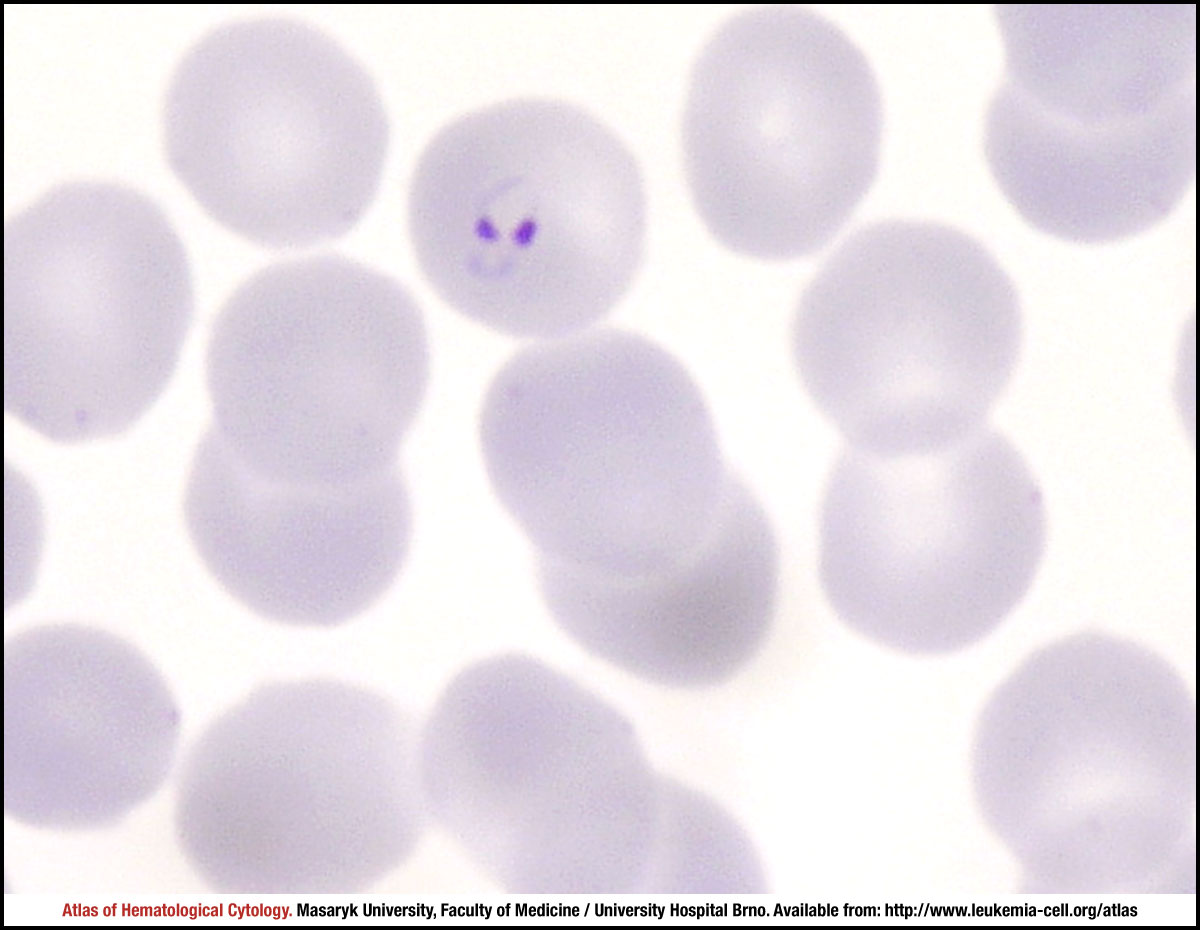 zoom picture
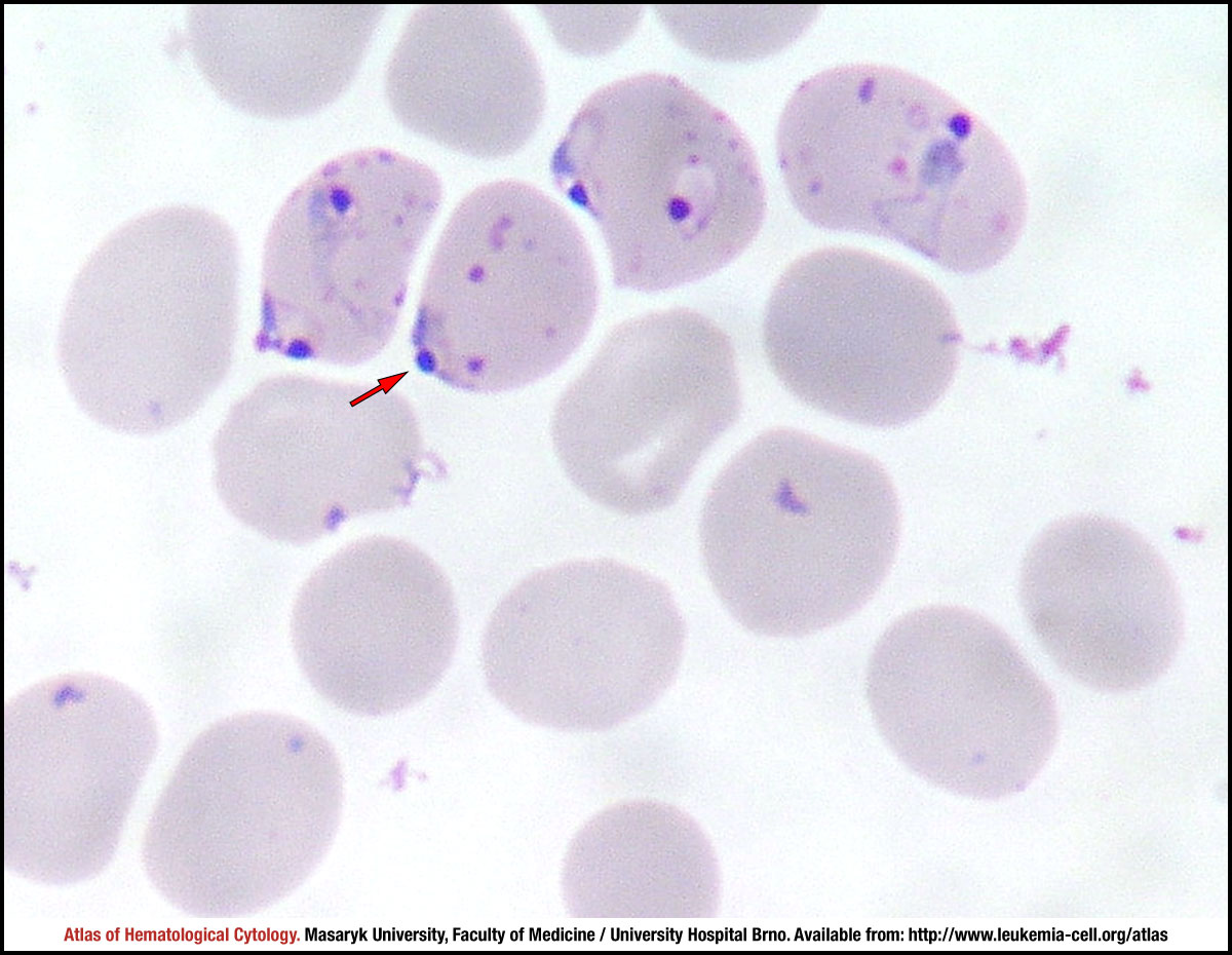 zoom picture
 zoom picture
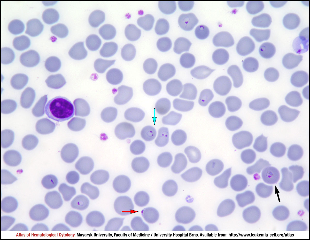 zoom picture
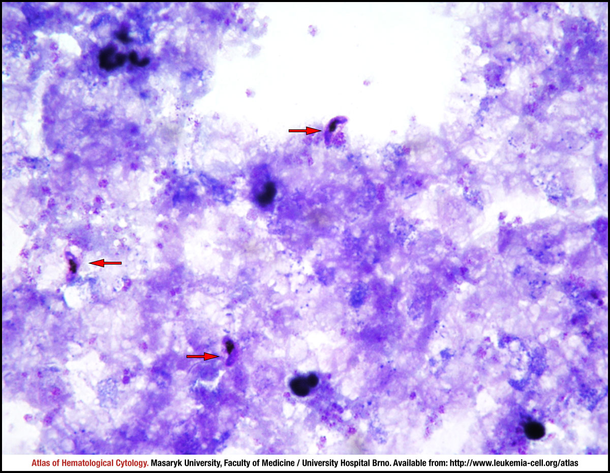 zoom picture
 zoom picture