with flowcytometry, cytogenetic and molecular biology findings
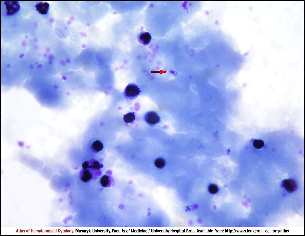
The red arrow shows a ring in a thick blood smear.
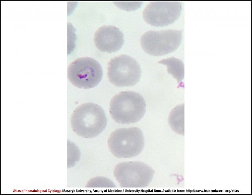
A ring in a thin blood smear. Infected erythrocytes are normal or smaller than normal in size, with normal shapes. The ring has a robust cytoplasm and a large chromatin dot.
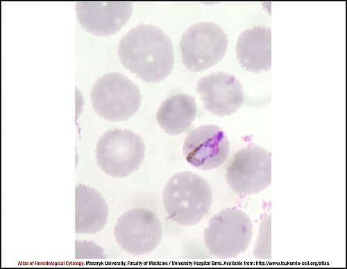
A trophozoite (band form) in a thin blood smear. Trophozoites have compact cytoplasm and are oval, round or band-shaped with dark-brown pigment. Erythrocytes infected with Plasmodium malariae have no stippling.
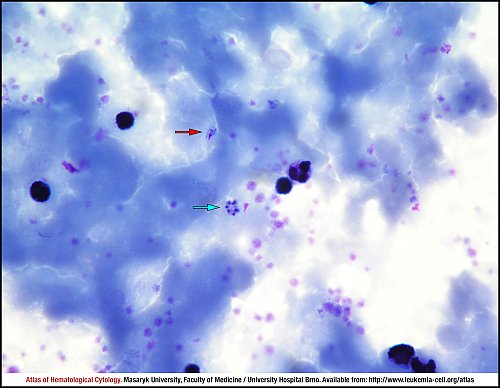
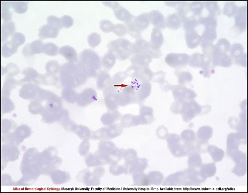
The red arrow marks a trophozoite, whereas the blue arrow shows a schizont in a thick blood smear. Plasmodium malariaeschizonts contain 6–12 merozoites, which can be arranged in a rosette or “daisy head” pattern.
The mature trophozoite shown in this thin blood smear is round to oval, has compact cytoplasm and dark-brown pigment.
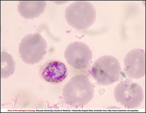
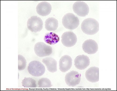
Schizonts in a thin blood smear. Merozoites are arranged around central pigment (the so-called “daisy head” pattern).
Schizonts in a thin blood smear. Merozoites are arranged around central pigment (the so-called “daisy head” pattern).
In this thin blood smear, the red arrow shows a schizont with merozoites arranged in a rosette pattern.
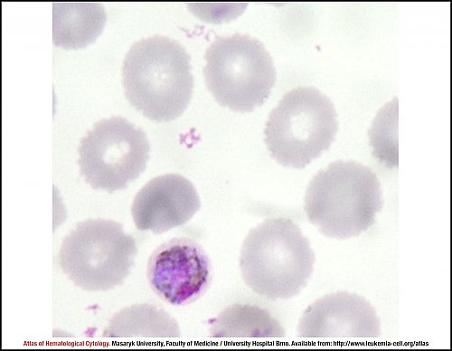
The mature macrogametocyte shown in this smear is round to slightly oval and almost fills the erythrocyte; blue plasma, small nucleus and brown pigment can be seen.
Atlas of Haematological Cytology [online]. 2016 [cit. 2026-4-02]. Available from WWW: http://www.leukemia-cell.org/atlas.
2026 CELL - Atlas of Haematological Cytology | site map
 zoom picture
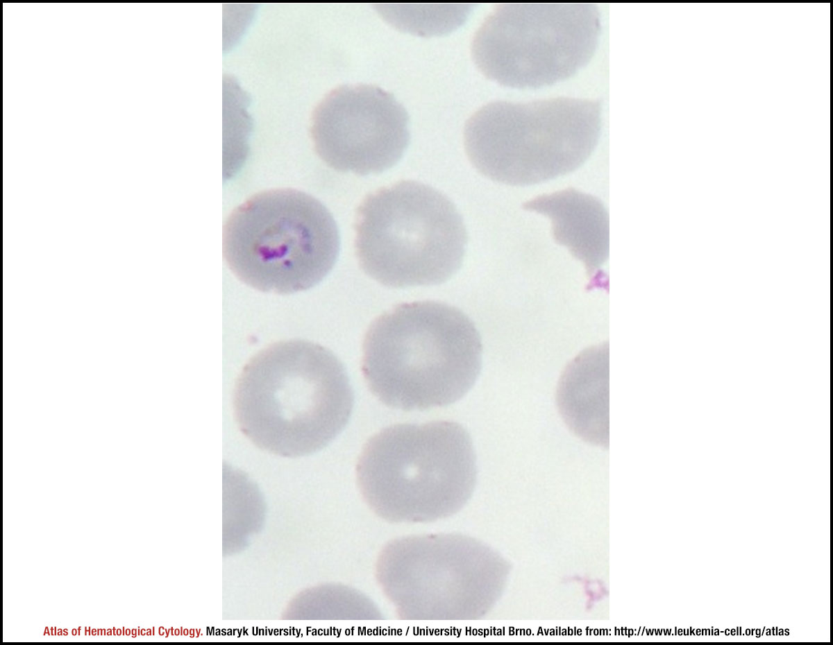 zoom picture
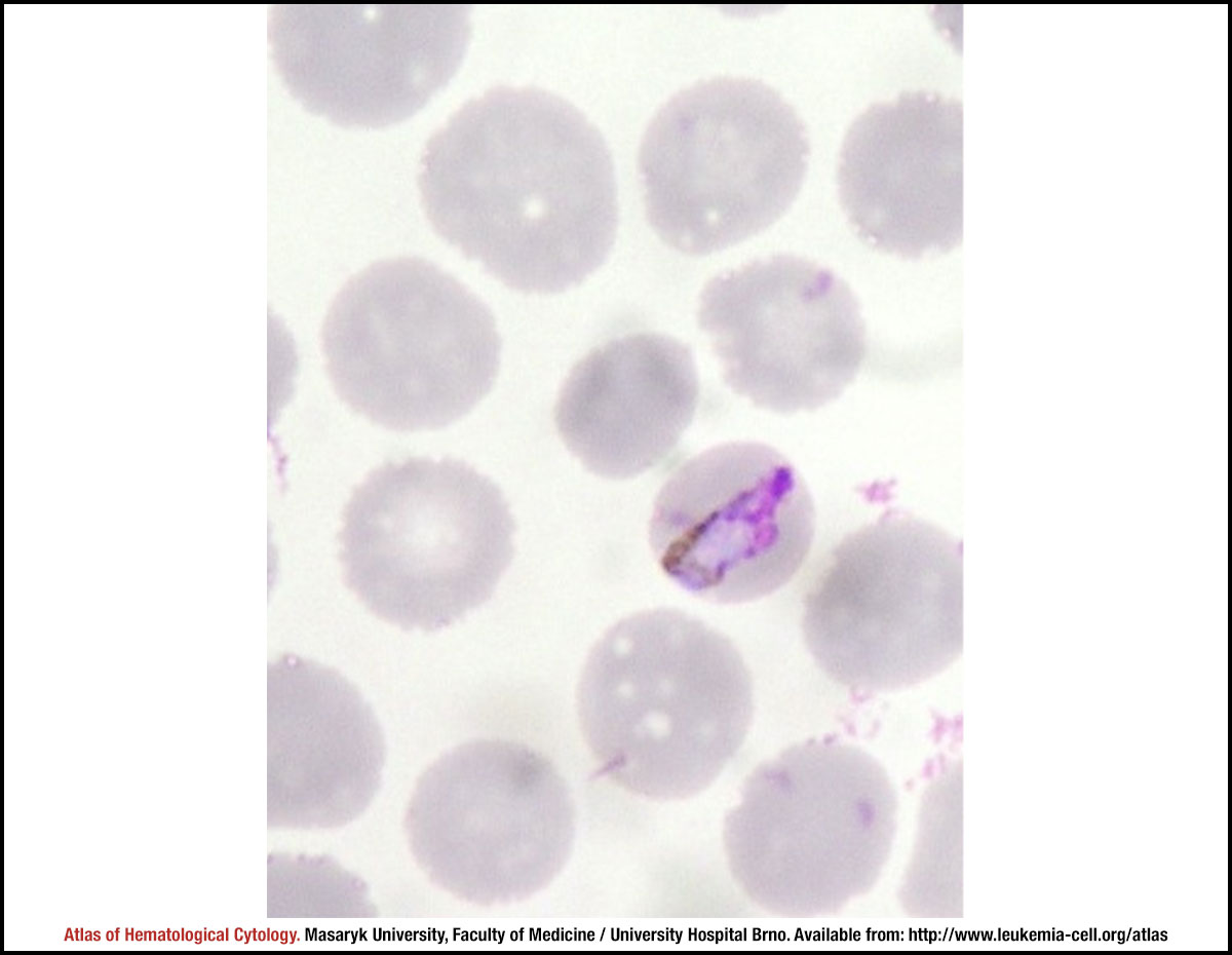 zoom picture
 zoom picture
 zoom picture
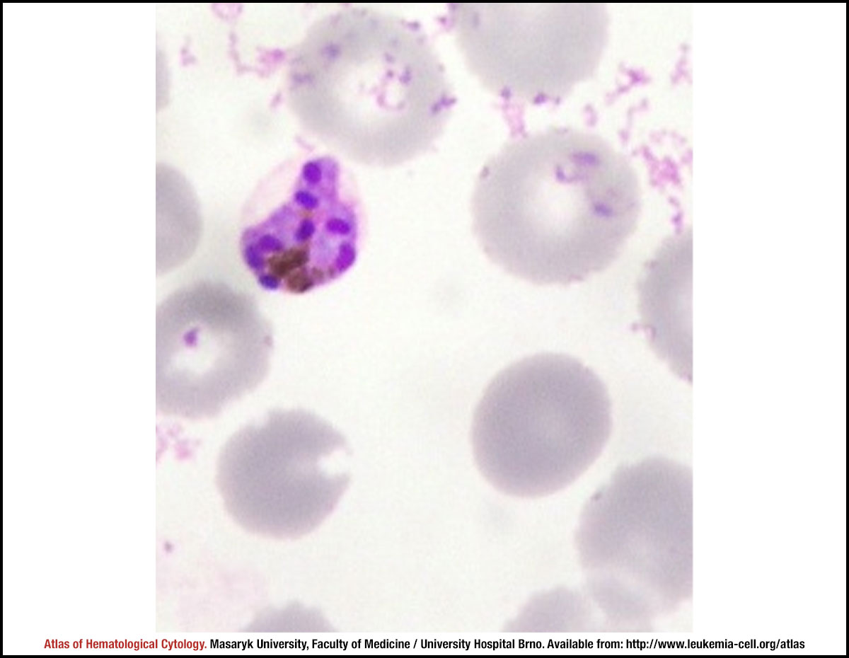 zoom picture
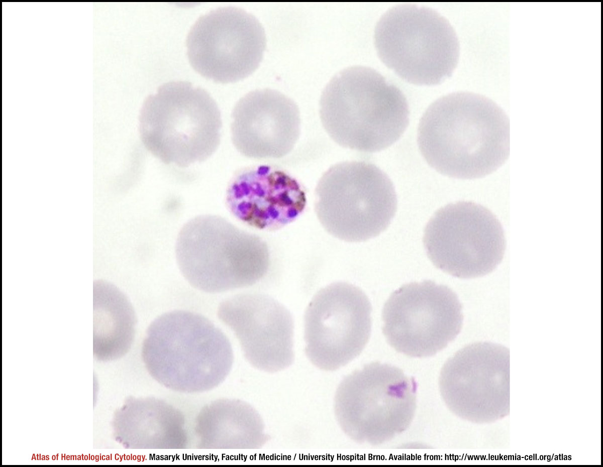 zoom picture
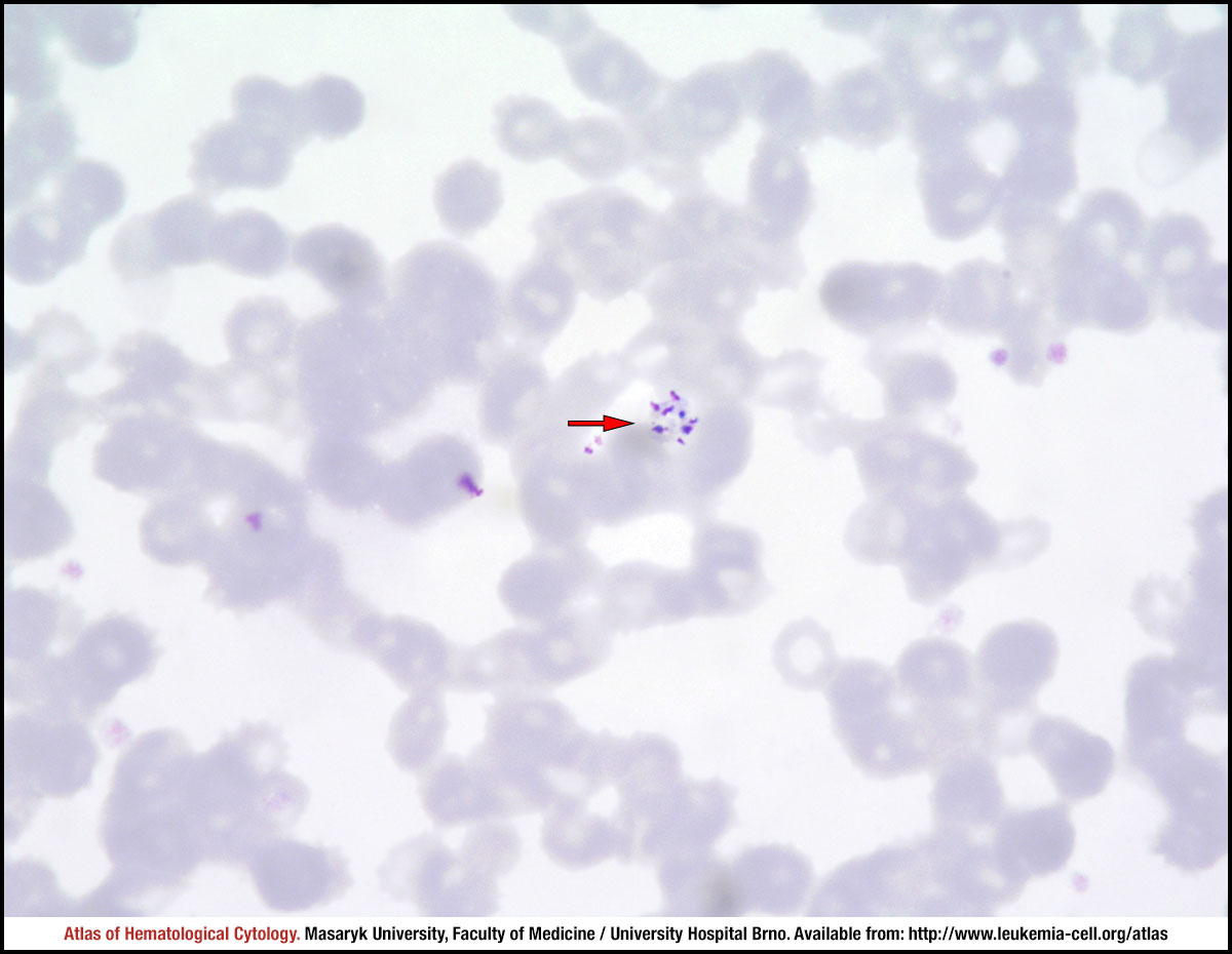 zoom picture
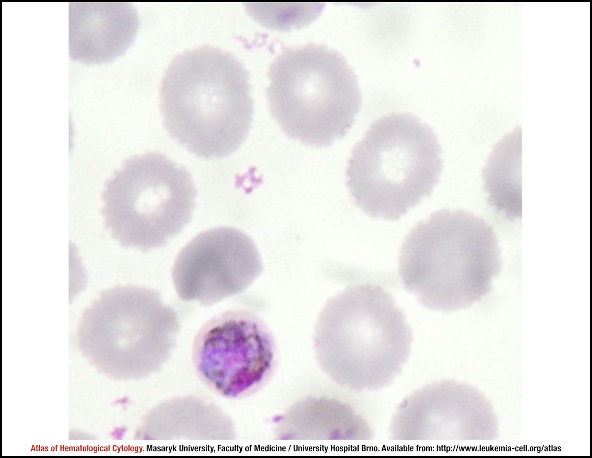 zoom picture