with flowcytometry, cytogenetic and molecular biology findings
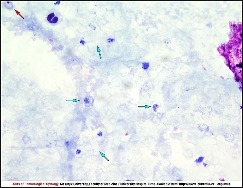
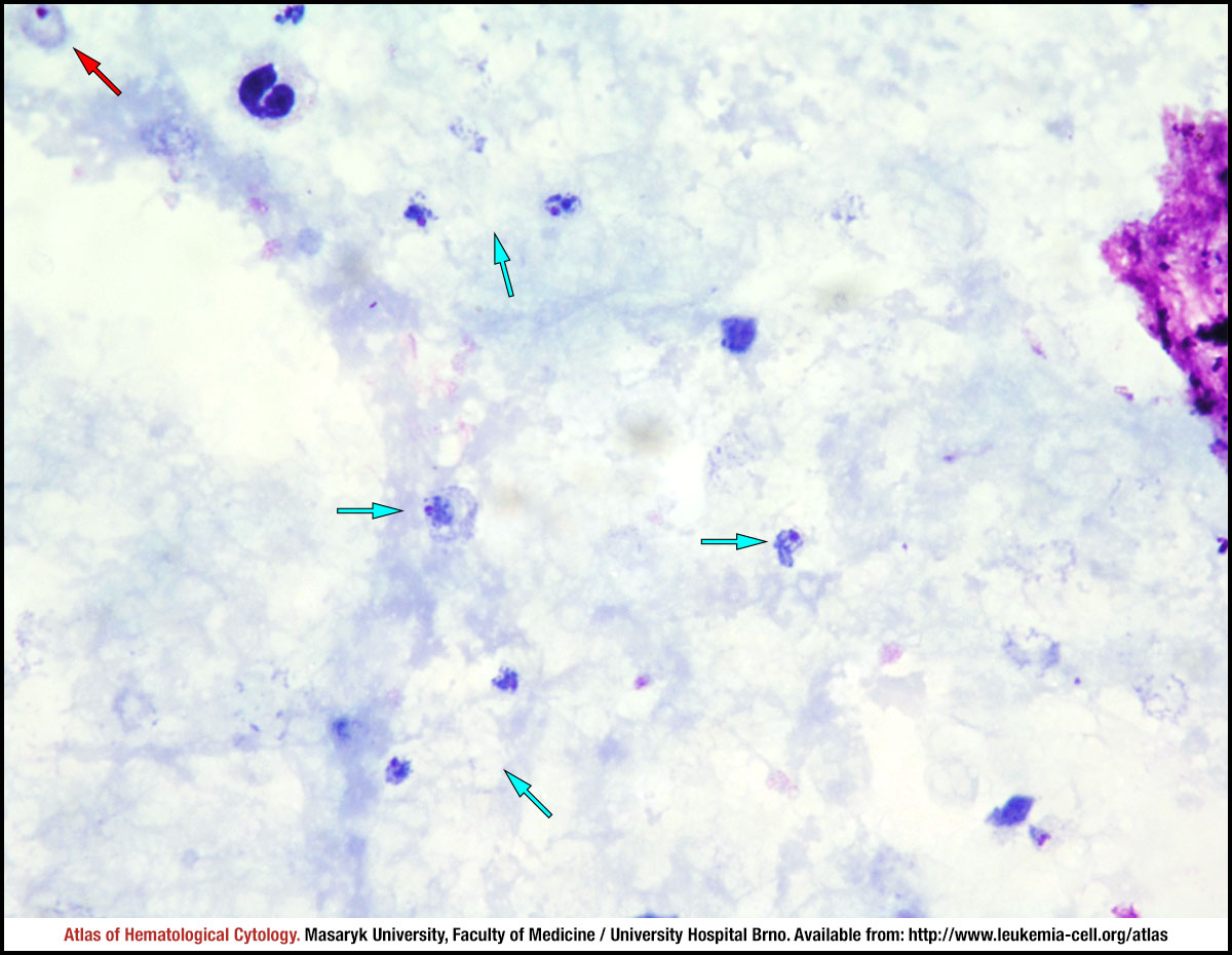
The red arrow shows a ring, whereas blue arrows mark trophozoites in this thick blood smear.
A ring in a thin blood smear. The infected erythrocyte is slightly oval-shaped and fimbriated, normal in size.
A ring in a thin blood smear. The infected erythrocyte is oval, enlarged and fimbriated.
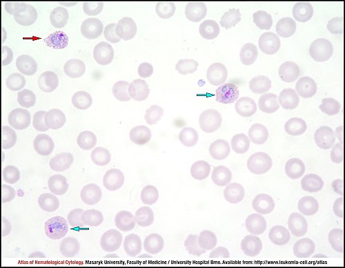
Several trophozoites are present in this thin blood smear. The red arrow shows a fimbriated trophozoite, whereas blue arrows mark compact to slightly irregular infected erythrocytes. James’ stippling is visible in all present trophozoites.
This developing trophozoite in a thin blood smear is slightly enlarged and oval-shaped and shows James’ stippling, which is darker than Schüffner’s dots and is usually present in all stages.
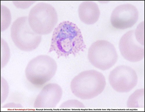
A Plasmodium ovale trophozoite with prominent fimbriation and James’ stippling in a thin blood smear.
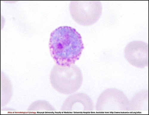
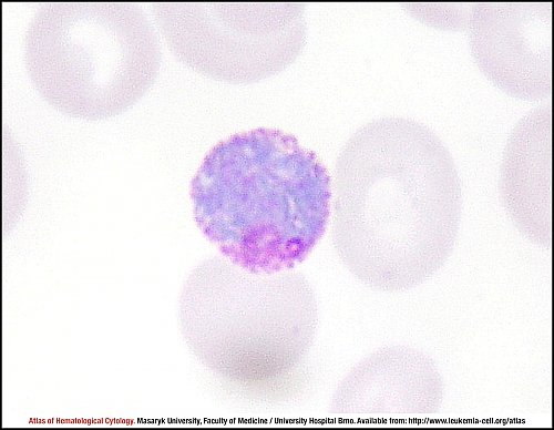
This gametocyte in a thin blood smear is round to oval and nearly fills the erythrocyte. Its red nucleus and James’ stippling also can be seen.
This gametocyte in a thin blood smear is round to oval and nearly fills the erythrocyte. Its red nucleus and James’ stippling also can be seen.
Atlas of Haematological Cytology [online]. 2016 [cit. 2024-4-26]. Available from WWW: http://www.leukemia-cell.org/atlas.
2024 CELL - Atlas of Haematological Cytology | site map
 zoom picture
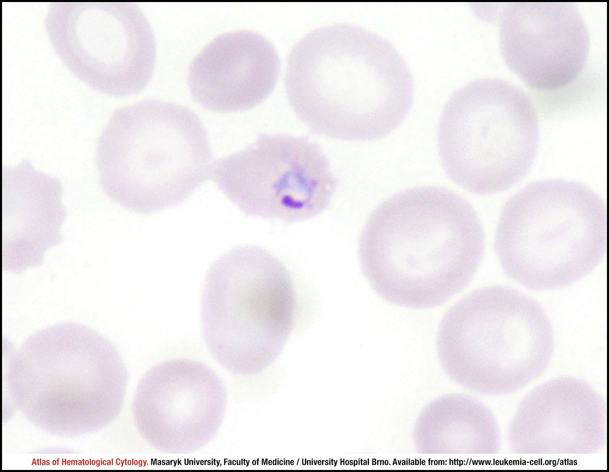 zoom picture
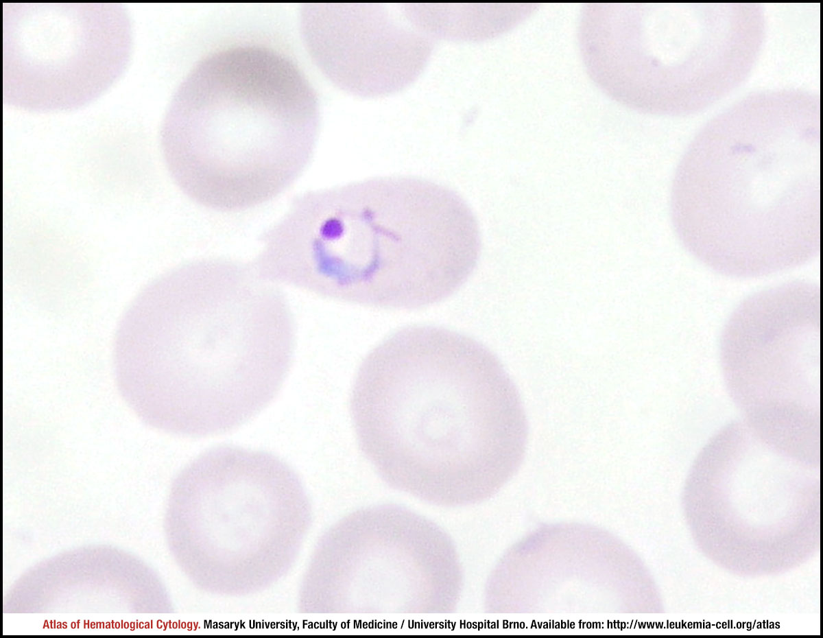 zoom picture
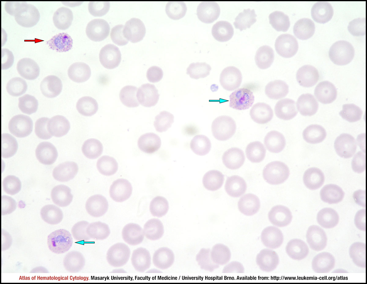 zoom picture
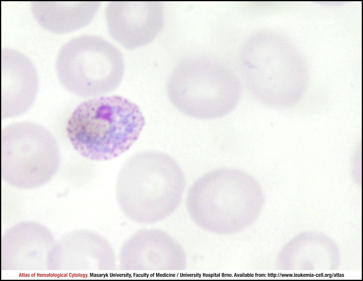 zoom picture
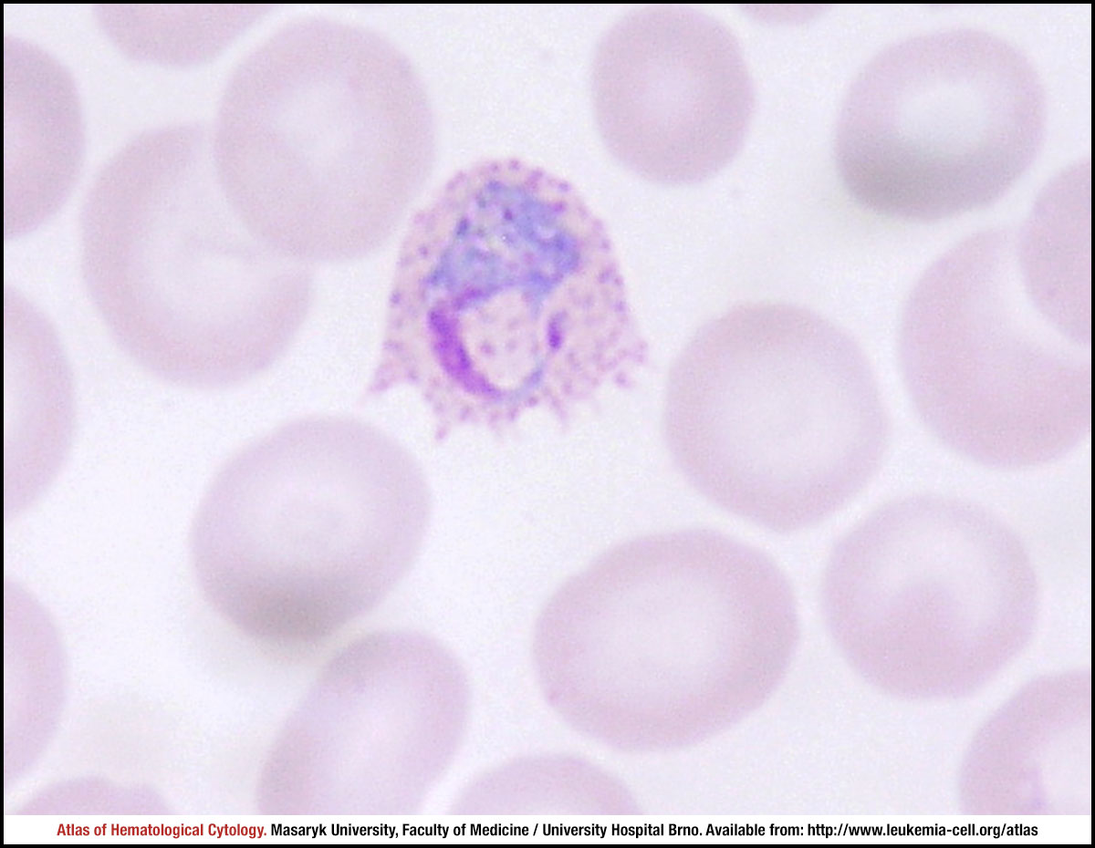 zoom picture
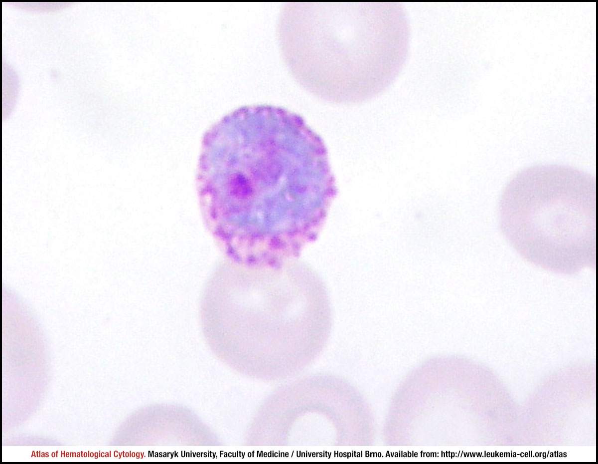 zoom picture
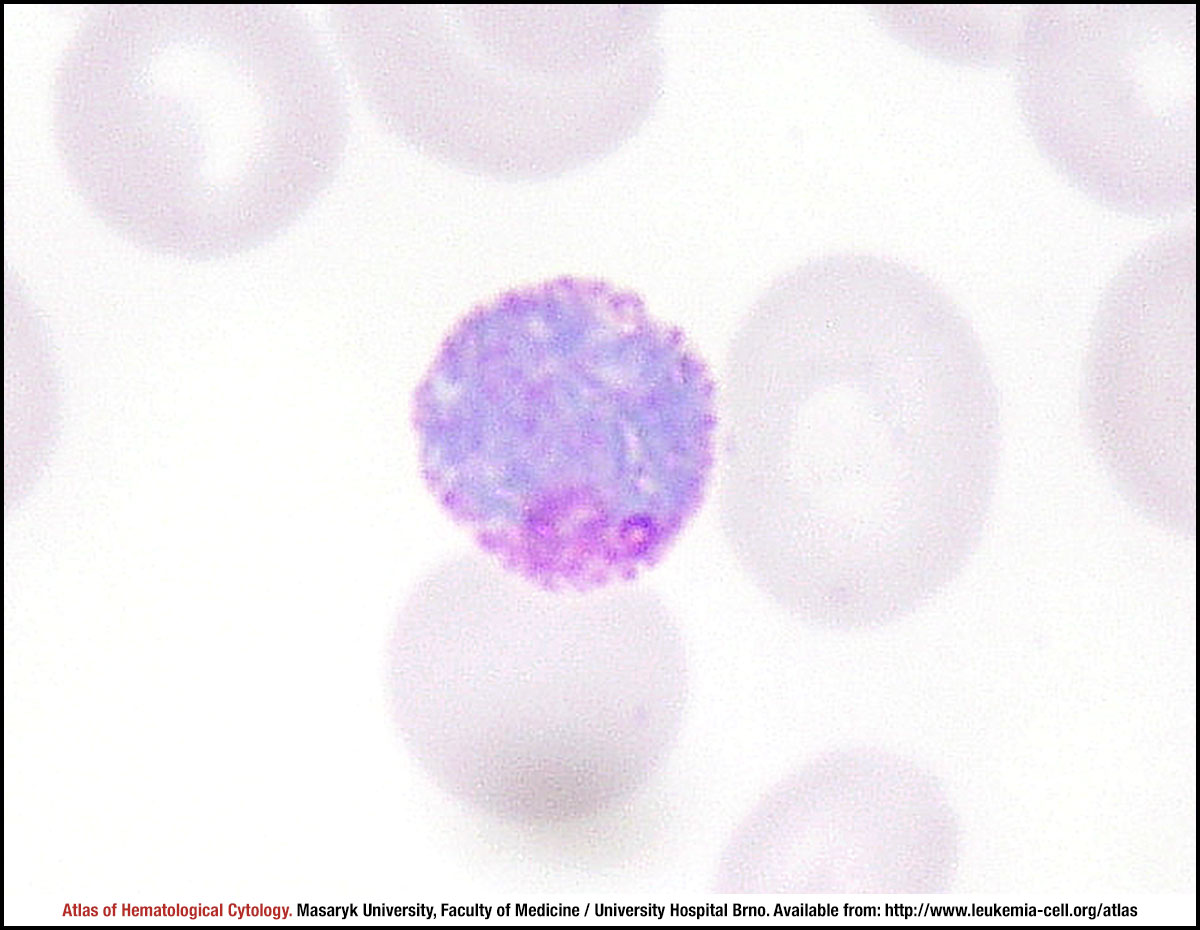 zoom picture