with flowcytometry, cytogenetic and molecular biology findings
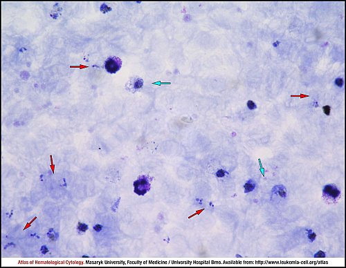
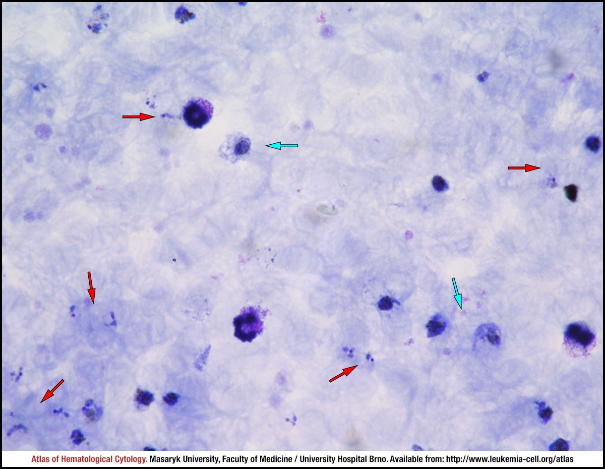
Red arrows show rings, whereas blue arrows show Plasmodium vivax trophozoites in a thick blood smear.
A thin blood smear of peripheral blood with erythrocytes infected with Plasmodium vivax. The red arrow marks a ring form, the erythrocyte is slightly enlarged and distorted. The blue arrow shows an amoeboid trophozoite, which is visibly enlarged and is “hugging” the neighbouring cell.
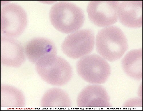
A young trophozoite (ring form) in a thin blood smear.
Growing trophozoites (amoeboid rings) in a thin blood smear. Infected erythrocytes are enlarged and distorted.
This thin blood smear shows a mature trophozoite. The infected erythrocyte is enlarged, distorted and Schüffner’s dots are visible. This Plasmodium vivax trophozoite resembles to band forms of Plasmodium malariae; the infected erythrocyte is up to twofold in size.
A growing trophozoite in a thin blood smear. The infected erythrocyte is enlarged, distorted and Schüffner’s dots are visible.
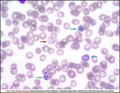
The red arrow shows an enlarged and “hugging” erythrocyte with an amoeboid ring. Blue arrows show almost mature gametocytes, which are enlarged, round to oval, have a scattered pigment and almost fill the infected erythrocytes. Thin blood smear.
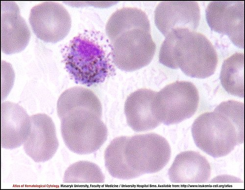
This macrogametocyte in a thin blood smear is round to oval, slightly enlarged. The parasite almost fills the infected erythrocyte. The cell has blue cytoplasm and eccentrically positioned nucleus, Schüffner’s dots and scattered brown pigment are visible.
This macrogametocyte in a thin blood smear is round to oval, slightly enlarged. The parasite almost fills the infected erythrocyte. The cell has blue cytoplasm and eccentrically positioned nucleus, Schüffner’s dots and scattered brown pigment are visible.
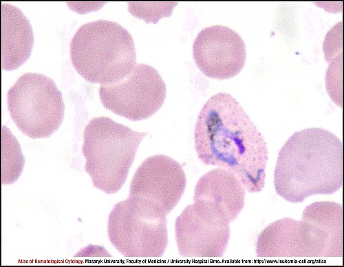
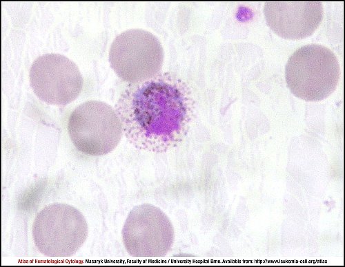
This mature microgametocyte has purple cytoplasm and bigger nucleus, brown pigment and Schüffner’s dots are also visible. An infected erythrocyte is enlarged and distorted.
The blue arrow shows a Plasmodium vivax macrogametocyte, whereas the red arrow marks a young immature schizont with yellowish-brown pigment in a thin blood smear.
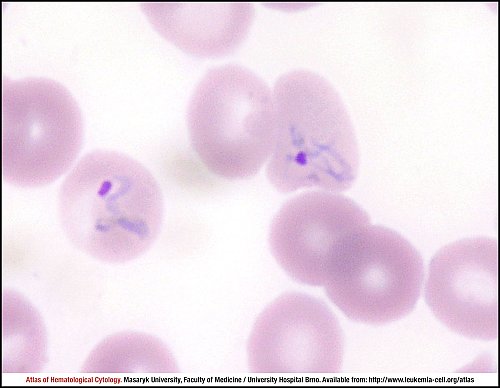
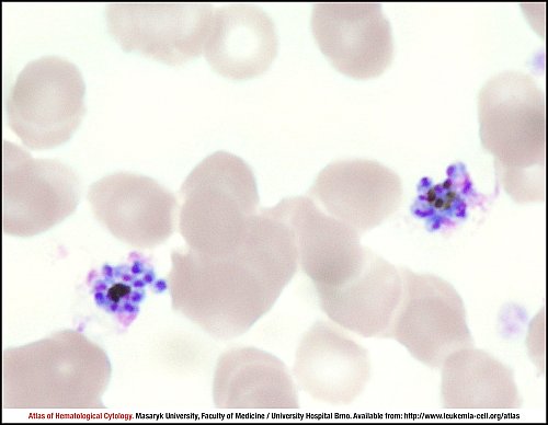
Plasmodium vivax schizonts in a thin blood smear are enlarged, contain 12 to 24 merozoites, usually fill the infected erythrocytes and show yellowish-brown pigment. Two mature schizonts are visible in this image.
Atlas of Haematological Cytology [online]. 2016 [cit. 2026-4-04]. Available from WWW: http://www.leukemia-cell.org/atlas.
2026 CELL - Atlas of Haematological Cytology | site map
 zoom picture
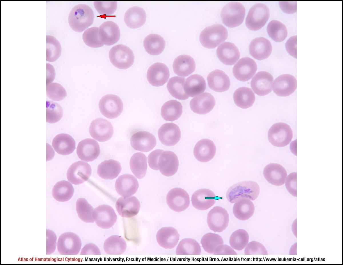 zoom picture
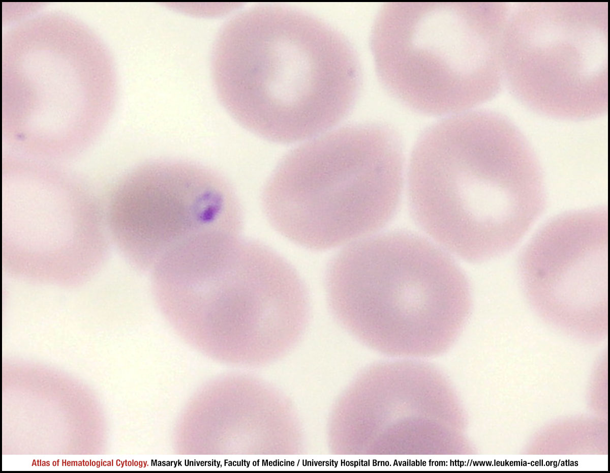 zoom picture
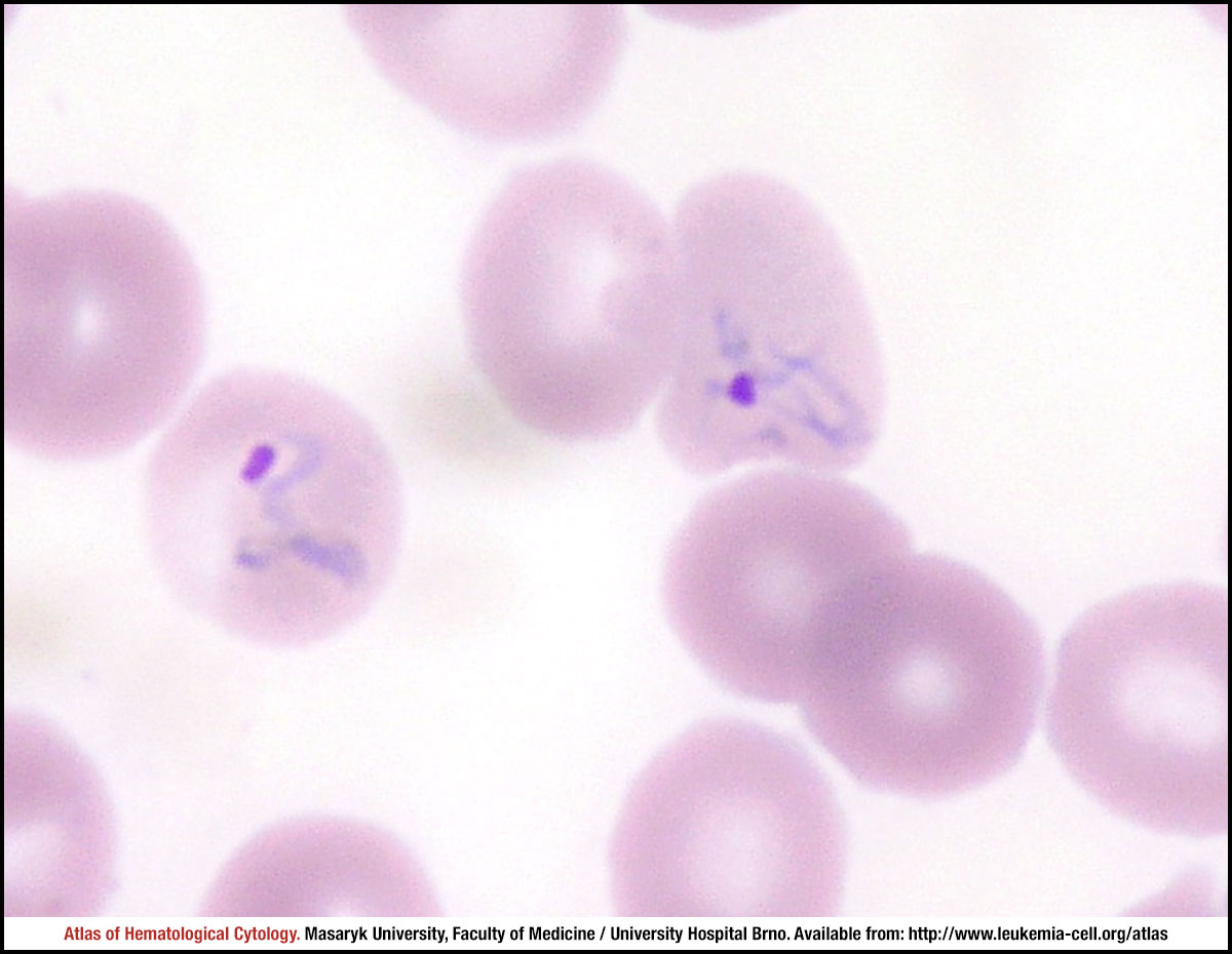 zoom picture
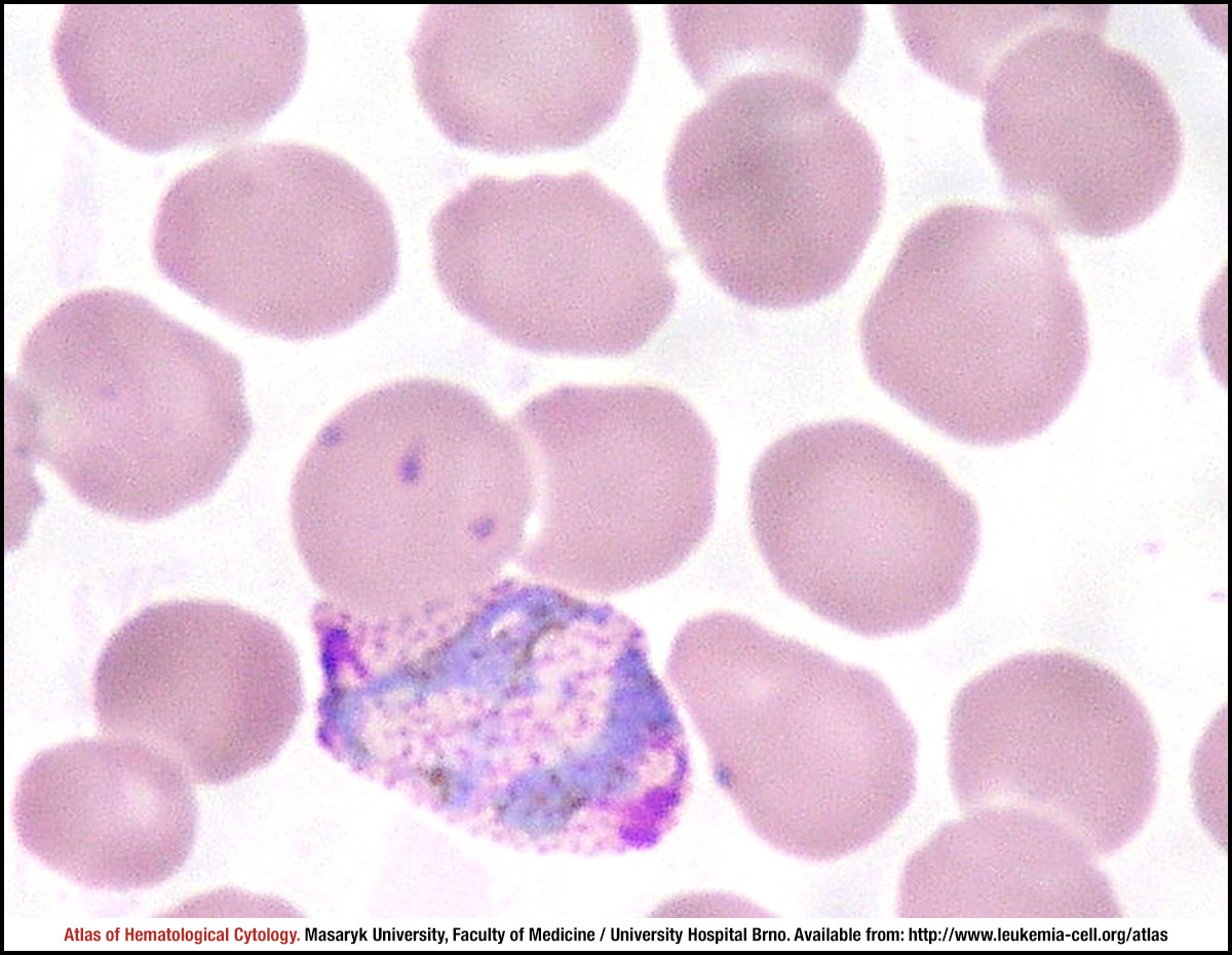 zoom picture
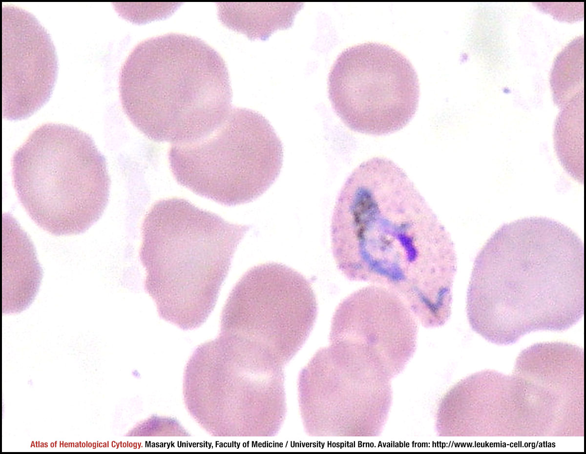 zoom picture
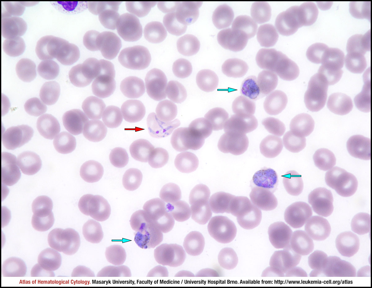 zoom picture
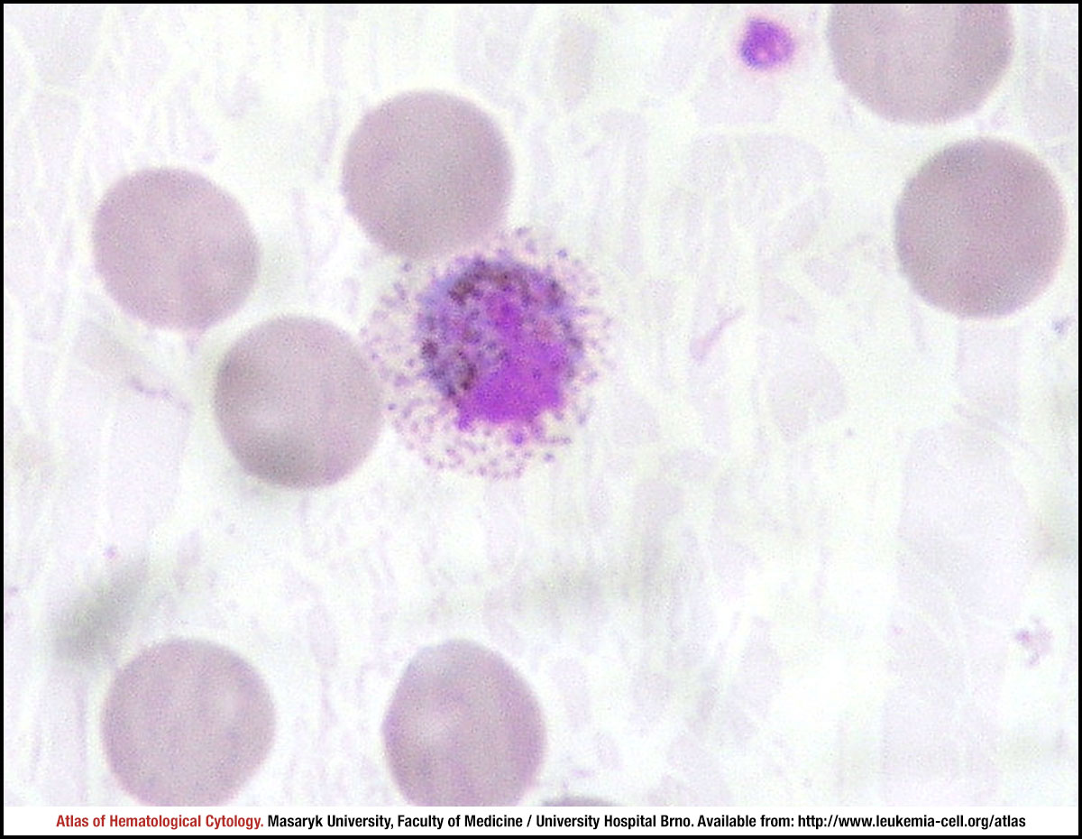 zoom picture
 zoom picture
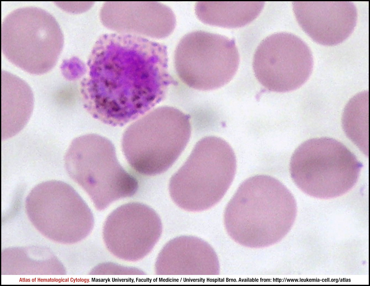 zoom picture
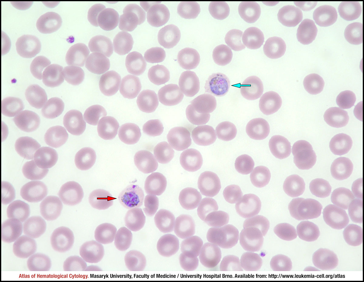 zoom picture
 zoom picture