with flowcytometry, cytogenetic and molecular biology findings
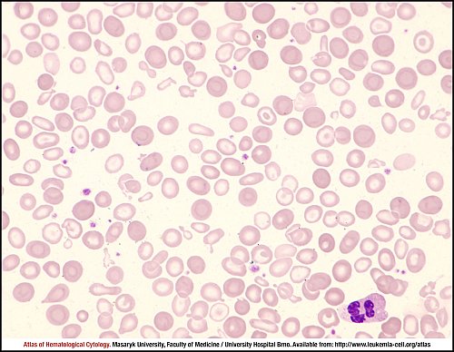
Marked anisopoikilocytosis, anisohypochromia and markedly atypical erythrocytes (in terms of shape) are shown in this peripheral blood smear. A segmented neutrophil is shown in the bottom right corner of the image.
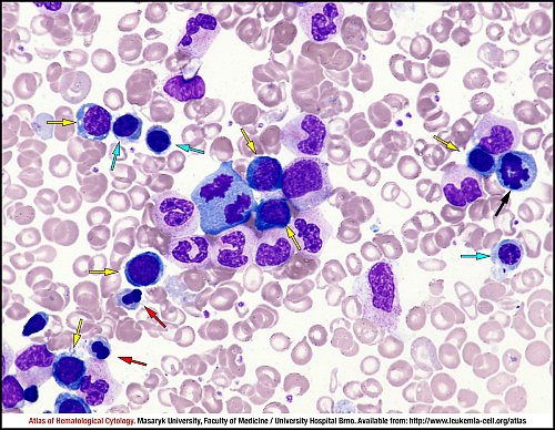
Basophilic, polychromatophilic and oxyphilic erythroblasts are marked by yellow, cyan and red arrows respectively. Cytoplasmic structure is disrupted in most of them (dehaemoglobinisation) and some of them have atypical shapes of nuclei (constrictions). A karyorrhectic figure of an erythroblast is shown by a black arrow. Other nuclear elements belong to morphologically adequate granulocytes and lymphocytes.
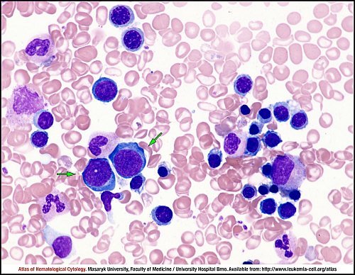
Proerythroblasts (i.e. the first developmental stage of erythropoiesis) are marked by green arrows. Otherwise, erythroblasts with impaired haemoglobinisation of cytoplasm, which is an indication of dyserythropoiesis, predominate in the image.
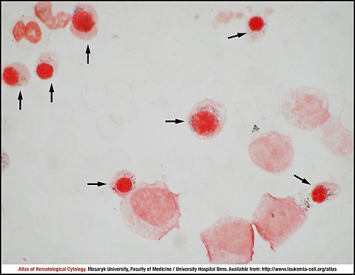
Iron stained marrow aspirate smear with many ring sideroblasts (black arrows), which are typical markers of sideroblastic dyserythropoesis. Other stained cells (on the top and on the bottom of the image) are granulocytes.
Atlas of Haematological Cytology [online]. 2016 [cit. 2024-4-25]. Available from WWW: http://www.leukemia-cell.org/atlas.
2024 CELL - Atlas of Haematological Cytology | site map
 zoom picture
 zoom picture
 zoom picture
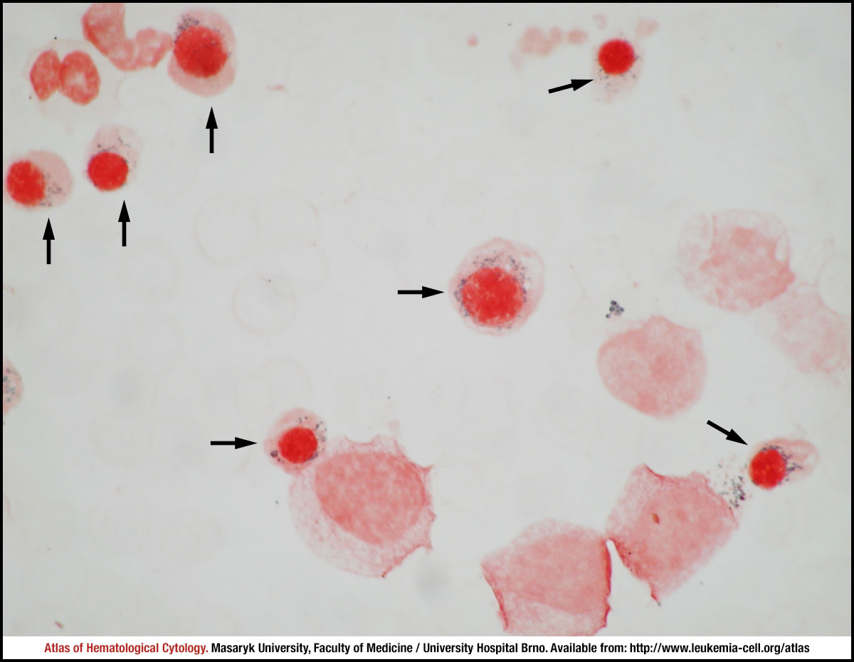 zoom picture