with flowcytometry, cytogenetic and molecular biology findings
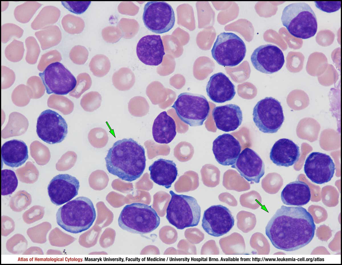
Peripheral blood smear with marked leukocytosis. The majority of cells are medium-sized lymphoid elements with round nuclei, visible nucleoli and relatively abundant and basophilic cytoplasm. Green arrows mark larger atypical prolymphocytes.
T-cell prolymphocytic leukaemia is characterised by an excessive leucocytosis and the presence of neoplastic prolymphocytes. Peripheral blood smear shows neoplastic prolymphocytes, which are medium-sized or large lymphoid cells with abundant basophilic cytoplasm, fine nuclear chromatin and prominent nucleoli (blue arrows). The shape of the nucleus is round or oval in a majority of neoplastic cells, but nuclear irregularities (red arrows) and anisocytosis are often visible, contrary to B-cell prolymphocytic leukaemia.
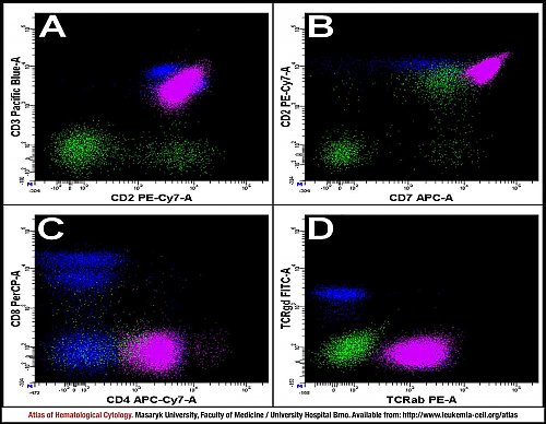
Analysis of a peripheral blood sample – colour marking of individual populations: neoplastic T lymphocytes (purple), T lymphocytes (blue), all lymphocytes (green), monocytes (yellow) and granulocytes (red).
Neoplastic T lymphocytes are characterised by the most commonly occurring immunophenotype in T-PLL: positivity for CD2, CD3, CD4, CD7 and TCR α/β and negativity for CD8 and TCR γ/δ (A–D).
Atlas of Haematological Cytology [online]. 2016 [cit. 2025-7-14]. Available from WWW: http://www.leukemia-cell.org/atlas.
2025 CELL - Atlas of Haematological Cytology | site map
 zoom picture
 zoom picture
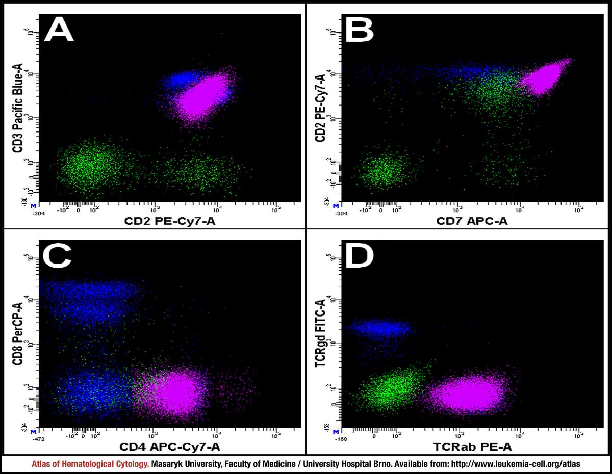 zoom picture